Gustave Roussy, Département de Chirurgie, Université Paris-Saclay, Villejuif, France.
Gustave Roussy, Plate-forme Imagerie et Cytométrie, UMS 23/3655, Université Paris-Saclay, Villejuif, France.
BJS Open. 2023 May 5;7(3). doi: 10.1093/bjsopen/zrad046.
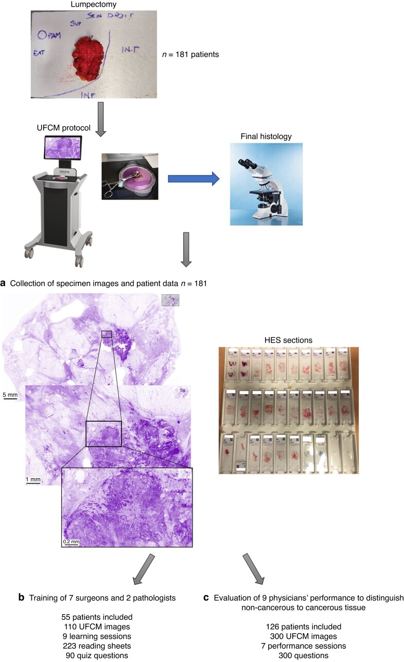
New generation ultra-fast fluorescence confocal microscopy allows the ex vivo intraoperative analysis of fresh tissue. The High resolution Imaging for Breast carcInoma detection in ex vivo Specimens after breast Conserving sUrgery by hiStolog Scanner (HIBISCUSS) project aimed to develop an online learning program to recognize the main breast tissue features on ultra-fast fluorescence confocal microscopy images and to evaluate the performance of surgeons and pathologists in diagnosing cancerous and non-cancerous breast tissue in ultra-fast fluorescence confocal microscopy images.
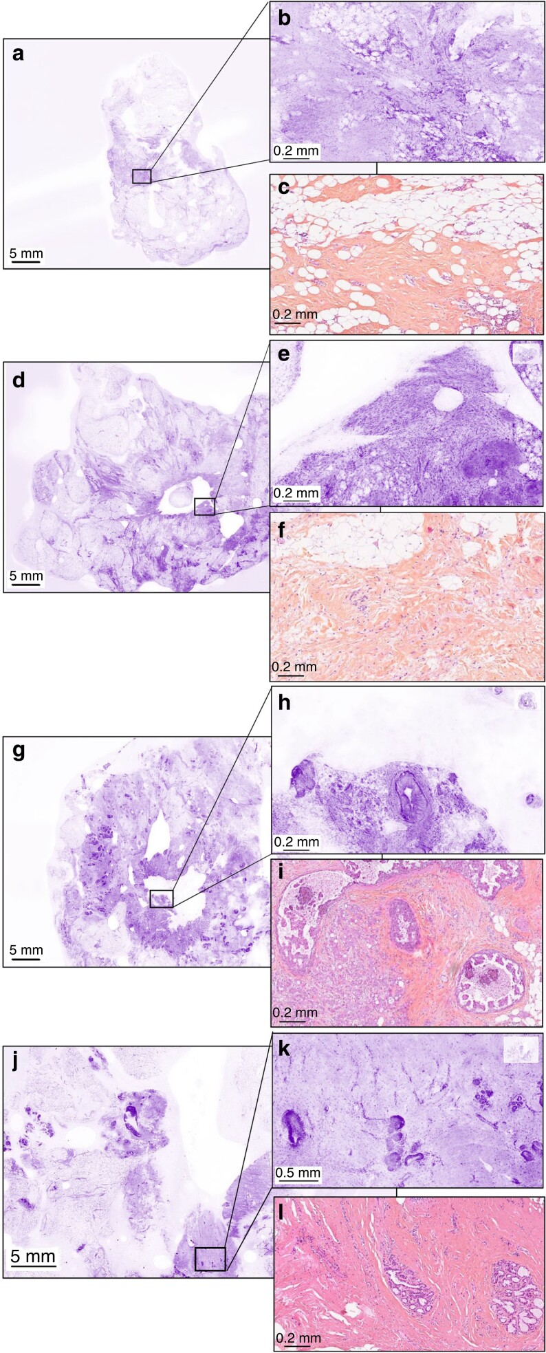
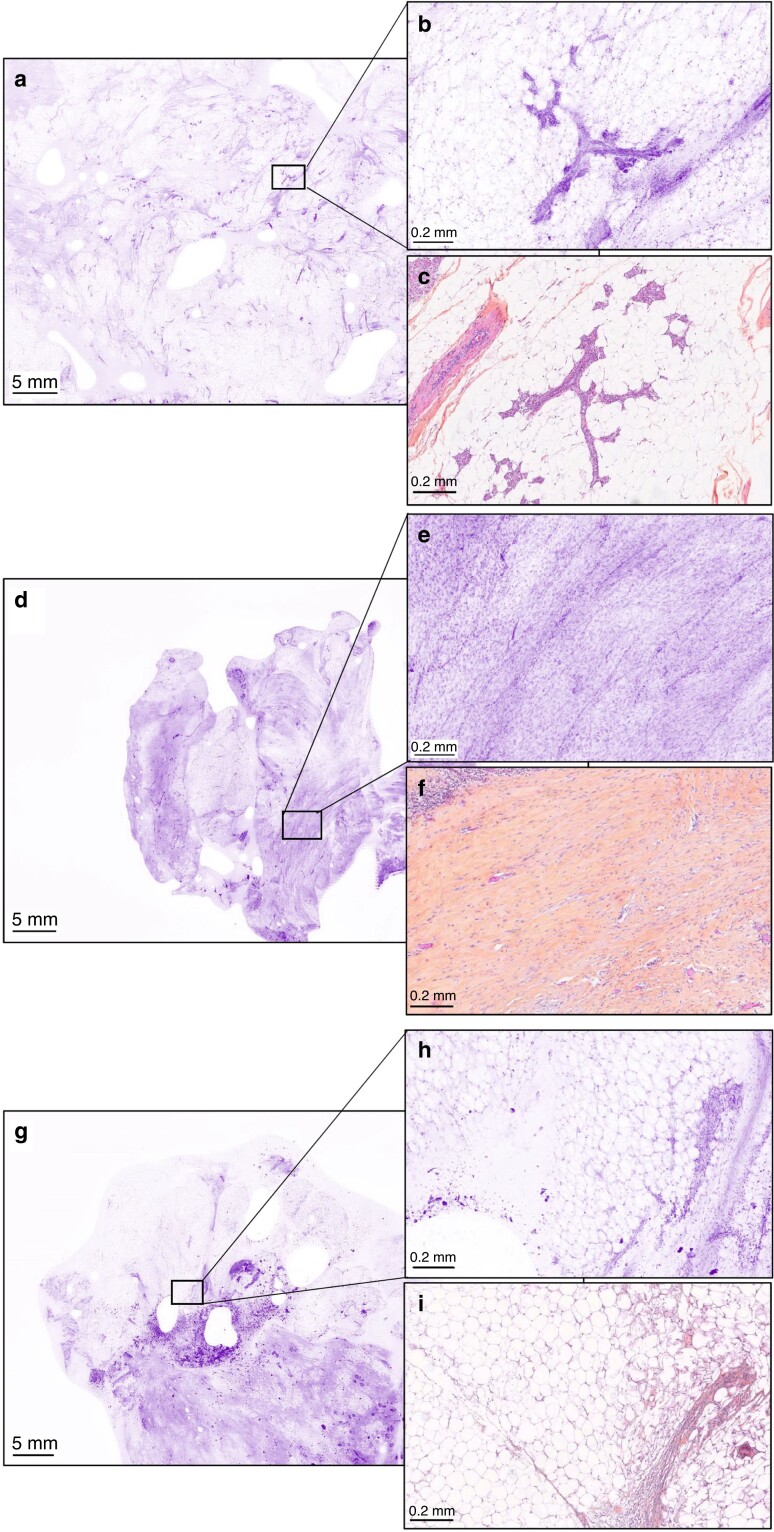
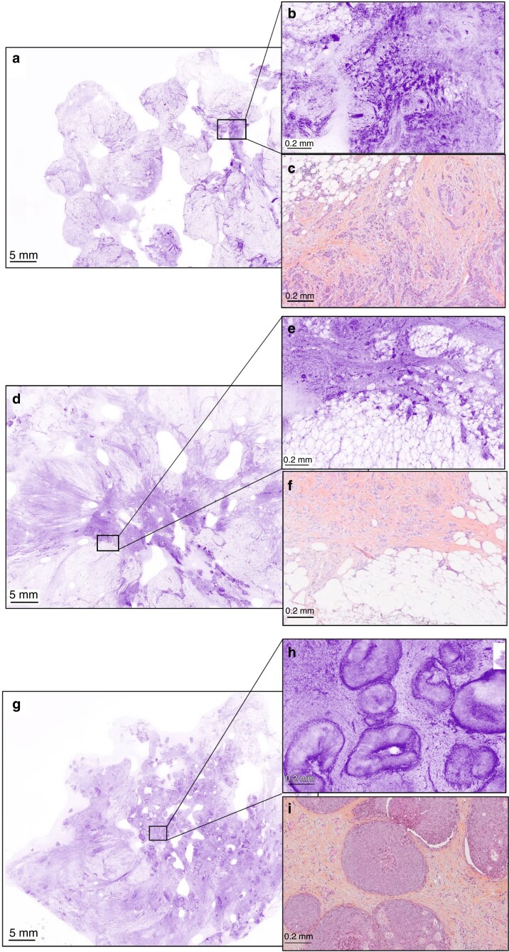
Patients who underwent conservative surgery or mastectomy for breast carcinoma (invasive or in situ lesions) were included. The fresh specimens were stained with a fluorescent dye and imaged using a large field-of-view (20 cm2) ultra-fast fluorescence confocal microscope.
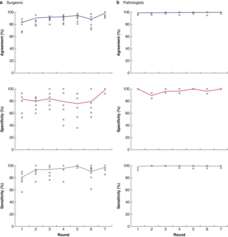
One hundred and eighty-one patients were included. The images from 55 patients were annotated to generate learning sheets and images from 126 patients were blindly interpreted by seven surgeons and two pathologists. The time for tissue processing and ultra-fast fluorescence confocal microscopy imaging was between 8 and 10 min. The training program was composed of 110 images divided into nine learning sessions. The final database for blind performance assessment comprised 300 images. The mean duration for one training session and one performance round was 17 and 27 min respectively. The performance of pathologists was almost perfect with 99.6 per cent (standard deviation (s.d.) 5.4 per cent) accuracy. Surgeons' accuracy significantly increased (P = 0.001) from 83 per cent (s.d. 8.4 per cent) in round 1 to 98 per cent (s.d. 4.1 per cent) in round 7 as well as the sensitivity (P = 0.004). Specificity increased without significance from 84 per cent (s.d. 16.7 per cent) in round 1 to 87 per cent (s.d. 16.4 per cent) in round 7 (P = 0.060).
Pathologists and surgeons showed a short learning curve in differentiating breast cancer from non-cancerous tissue in ultra-fast fluorescence confocal microscopy images. Performance assessment for both specialties supports ultra-fast fluorescence confocal microscopy evaluation for intraoperative management.
NCT04976556 (http://www.clinicaltrials.gov).
新一代超快速荧光共聚焦显微镜可实现新鲜组织的离体术中分析。通过组织扫描高分辨率成像在保乳手术后检测乳腺肿瘤的研究(HIBISCUSS)项目旨在开发一个在线学习程序,以识别超快速荧光共聚焦显微镜图像上的主要乳腺组织特征,并评估外科医生和病理学家在诊断超快速荧光共聚焦显微镜图像中癌性和非癌性乳腺组织的性能。
纳入接受保乳手术或乳房切除术治疗的乳腺癌(浸润性或原位病变)患者。新鲜标本用荧光染料染色,使用大视场(20 cm2)超快速荧光共聚焦显微镜进行成像。
共纳入 181 例患者。55 例患者的图像进行注释以生成学习表,126 例患者的图像由 7 名外科医生和 2 名病理学家进行盲法解读。组织处理和超快速荧光共聚焦显微镜成像时间为 8 至 10 分钟。培训计划由 110 张图像组成,分为九个学习课程。盲法性能评估的最终数据库包含 300 张图像。一个学习课程和一轮性能评估的平均持续时间分别为 17 分钟和 27 分钟。病理学家的表现几乎完美,准确率为 99.6%(标准差为 5.4%)。外科医生的准确性显著提高(P = 0.001),从第一轮的 83%(标准差为 8.4%)提高到第七轮的 98%(标准差为 4.1%),以及敏感性(P = 0.004)。特异性从第一轮的 84%(标准差为 16.7%)提高到第七轮的 87%(标准差为 16.4%),但无统计学意义(P = 0.060)。
病理学家和外科医生在超快速荧光共聚焦显微镜图像中区分乳腺癌和非癌性组织的学习曲线较短。对这两个专业的性能评估均支持超快速荧光共聚焦显微镜用于术中管理。
NCT04976556(http://www.clinicaltrials.gov)。