Comprehensive Heart Failure Center (CHFC), Chair of Molecular and Cellular Imaging, University Hospital Würzburg, Würzburg, Germany.
Department of Diagnostic and Interventional Radiology, University Hospital Würzburg, Würzburg, Germany.
PLoS One. 2022 Jun 29;17(6):e0270689. doi: 10.1371/journal.pone.0270689. eCollection 2022.
To investigate the effects of B1-shimming and radiofrequency (RF) parallel transmission (pTX) on the visualization and quantification of the degree of stenosis in a coronary artery phantom using 7 Tesla (7 T) magnetic resonance imaging (MRI).
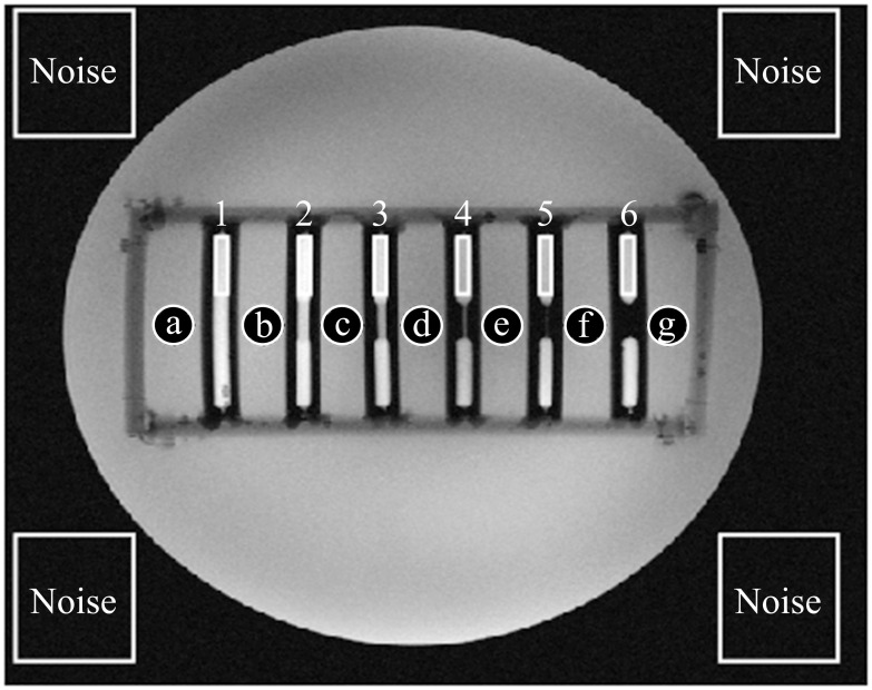
Stenosis phantoms with different grades of stenosis (0%, 20%, 40%, 60%, 80%, and 100%; 5 mm inner vessel diameter) were produced using 3D printing (clear resin). Phantoms were imaged with four different concentrations of diluted Gd-DOTA representing established arterial concentrations after intravenous injection in humans. Samples were centrally positioned in a thorax phantom of 30 cm diameter filled with a custom-made liquid featuring dielectric properties of muscle tissue. MRI was performed on a 7 T whole-body system. 2D-gradient-echo sequences were acquired with an 8-channel transmit 16-channel receive (8 Tx / 16 Rx) cardiac array prototype coil with and without pTX mode. Measurements were compared to those obtained with identical scan parameters using a commercially available 1 Tx / 16 Rx single transmit coil (sTX). To assess reproducibility, measurements (n = 15) were repeated at different horizontal angles with respect to the B0-field.
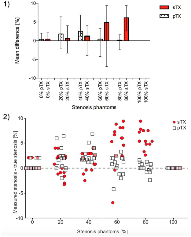
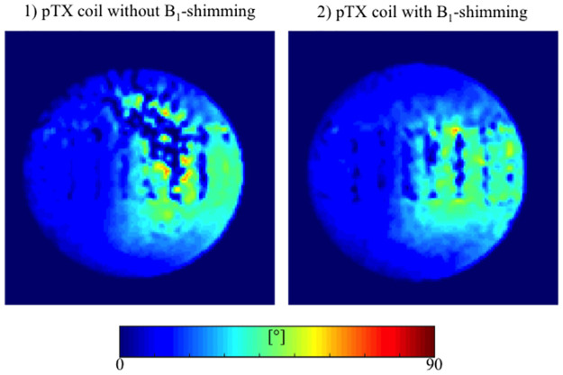
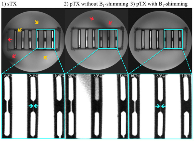
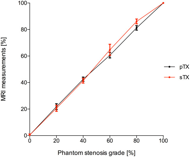
B1-shimming and pTX markedly improved flip angle homogeneity across the thorax phantom yielding a distinctly increased signal-to-noise ratio (SNR) averaged over a whole slice relative to non-manipulated RF fields. Images without B1-shimming showed shading artifacts due to local B1+-field inhomogeneities, which hampered stenosis quantification in severe cases. In contrast, B1-shimming and pTX provided superior image homogeneity. Compared with a conventional sTX coil higher grade stenoses (60% and 80%) were graded significantly (p<0.01) more precise. Mild to moderate grade stenoses did not show significant differences. Overall, SNR was distinctly higher with B1-shimming and pTX than with the conventional sTX coil (inside the stenosis phantoms 14%, outside the phantoms 32%). Both full and half concentration (10.2 mM and 5.1 mM) of a conventional Gd-DOTA dose for humans were equally suitable for stenosis evaluation in this phantom study.
B1-shimming and pTX at 7 T can distinctly improve image homogeneity and therefore provide considerably more accurate MR image analysis, which is beneficial for imaging of small vessel structures.
本研究旨在探讨在 7 特斯拉(7T)磁共振成像(MRI)中,B1 匀场和射频(RF)并行传输(pTX)对冠状动脉模型狭窄程度可视化和定量的影响。
使用 3D 打印(透明树脂)制作了不同狭窄程度(0%、20%、40%、60%、80%和 100%;内血管直径 5 毫米)的狭窄模型。在一个直径为 30 厘米的胸腔模型中,使用四个不同浓度的稀释 Gd-DOTA 进行成像,这些浓度代表了人体静脉注射后的动脉浓度。样品放置在充满定制液体的胸腔模型中央,该液体具有肌肉组织的介电特性。在一台 7T 全身系统上进行 MRI。使用 8 通道发射 16 通道接收(8Tx/16Rx)心脏阵列原型线圈,采集具有和不具有 pTX 模式的二维梯度回波序列。与使用商业上可用的 1Tx/16Rx 单发射线圈(sTX)获得的相同扫描参数的测量结果进行了比较。为了评估可重复性,使用不同水平角度相对于 B0 场重复测量(n=15)。
B1 匀场和 pTX 显著改善了整个胸腔模型的翻转角均匀性,与未经处理的 RF 场相比,整个切片的信号噪声比(SNR)明显增加。未经 B1 匀场的图像由于局部 B1+场不均匀性而出现阴影伪影,这使得严重情况下的狭窄定量变得困难。相比之下,B1 匀场和 pTX 提供了更好的图像均匀性。与传统的 sTX 线圈相比,更高程度的狭窄(60%和 80%)分级明显更准确(p<0.01)。轻度至中度程度的狭窄没有显著差异。总体而言,B1 匀场和 pTX 比传统的 sTX 线圈具有明显更高的 SNR(在狭窄模型内部 14%,在模型外部 32%)。对于人体的常规 Gd-DOTA 剂量的全浓度(10.2mM)和半浓度(5.1mM)对于这种在模型研究中的狭窄评估同样适用。
在 7T 时,B1 匀场和 pTX 可以明显改善图像均匀性,从而提供更准确的磁共振图像分析,这对于小血管结构的成像非常有益。