Deaver J William, Schrems Eleanor R, Brown Lemuel A, Haynie Wesley A, Perry Richard A, Rosa-Caldwell Megan E, Tedrowe Michelle A, Greene Nicholas P, Washington Tyrone A
Cachexia Research Laboratory, Exercise Science Research Center, Department of Health, Human Performance and Recreation, University of Arkansas, Fayetteville, AR, 72701, USA.
Exercise Muscle Biology Laboratory, Exercise Science Research Center, Department of Health, Human Performance and Recreation, University of Arkansas, Fayetteville, AR, 72701, USA.
Sports Med Health Sci. 2021 Oct 12;3(4):212-217. doi: 10.1016/j.smhs.2021.09.003. eCollection 2021 Dec.
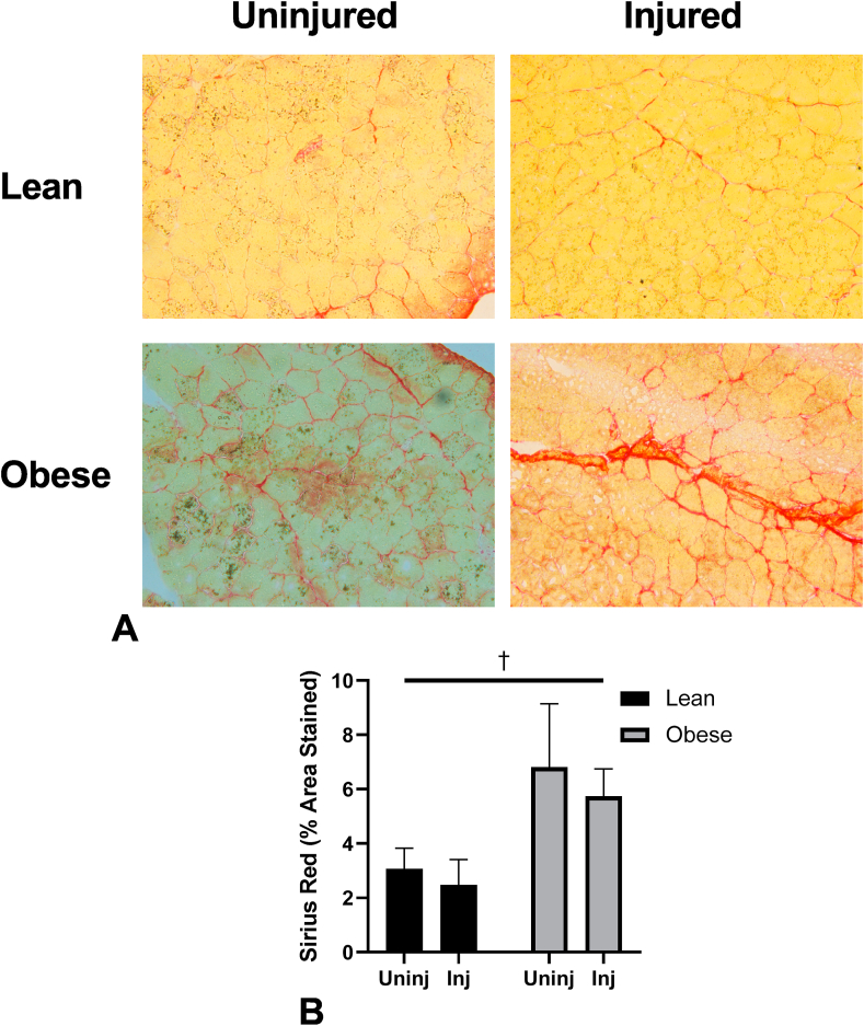
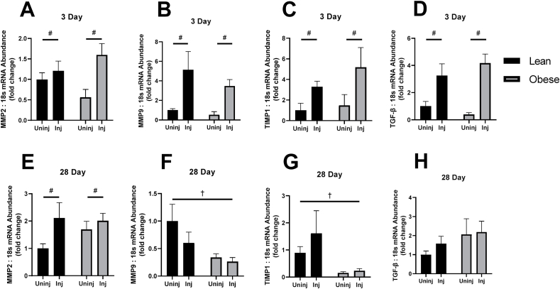
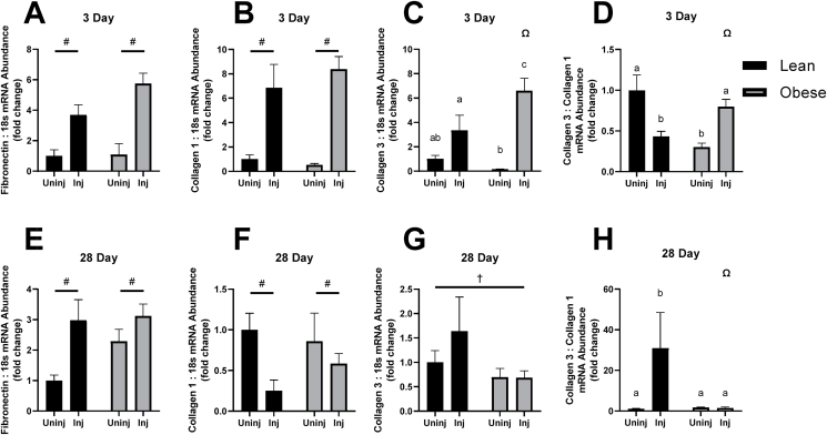
Diet-induced obesity has previously been shown to occur with the concomitant rise in the expression of proinflammatory cytokines and increases in collagen deposition. While it has been known that the regenerative process of skeletal muscle is altered in obese mice following an acute muscle injury, we sought to examine differences in the expression of various markers of extracellular matrix remodeling and repair. Our laboratory has previously reported an impaired inflammatory and protein synthetic signaling in these mice that may contribute negatively to the muscle regenerative process. To expand upon this previous investigation, tissues from these animals underwent further analysis to determine the extent of changes to the regenerative response within the extracellular matrix, including transcriptional changes in Collagen I, Collagen III, and Fibronectin. Here, we show that the expression of Collagen III:I is significantly increased at 3-days post-injury in obese injured animals compared to lean injured animals ( = 0.0338), and by 28-days the obese injured animals exhibit a significantly lower Collagen III:I than their lean injured counterparts ( = 0.0035). We demonstrate an impaired response to an acute muscle injury in obese mice when compared with lean counterparts. However, further studies are required to elucidate translational consequences of these changes, as well as to determine any causative mechanisms that may be driving this effect.
先前的研究表明,饮食诱导的肥胖伴随着促炎细胞因子表达的增加和胶原蛋白沉积的增多。虽然已知肥胖小鼠在急性肌肉损伤后骨骼肌的再生过程会发生改变,但我们试图研究细胞外基质重塑和修复的各种标志物表达的差异。我们实验室先前报道过这些小鼠存在炎症和蛋白质合成信号受损的情况,这可能对肌肉再生过程产生负面影响。为了扩展之前的研究,对这些动物的组织进行了进一步分析,以确定细胞外基质内再生反应的变化程度,包括I型胶原蛋白、III型胶原蛋白和纤连蛋白的转录变化。在此,我们表明,与瘦损伤动物相比,肥胖损伤动物在损伤后3天时III型胶原蛋白与I型胶原蛋白的表达显著增加(P = 0.0338),到28天时,肥胖损伤动物的III型胶原蛋白与I型胶原蛋白表达显著低于瘦损伤动物(P = 0.0035)。我们证明,与瘦小鼠相比,肥胖小鼠对急性肌肉损伤的反应受损。然而,需要进一步研究来阐明这些变化的转化后果,以及确定可能导致这种效应的任何致病机制。