Xu Pengfei, Werner Jens-Uwe, Milerski Sebastian, Hamp Carmen M, Kuzenko Tatjana, Jähnert Markus, Gottmann Pascal, de Roy Luisa, Warnecke Daniela, Abaei Alireza, Palmer Annette, Huber-Lang Markus, Dürselen Lutz, Rasche Volker, Schürmann Annette, Wabitsch Martin, Knippschild Uwe
Department of General and Visceral Surgery, Ulm University Hospital, Ulm, Germany.
Department of Experimental Diabetology, German Institute of Human Nutrition, Potsdam-Rehbrücke, Potsdam, Germany.
Front Physiol. 2018 Jun 5;9:674. doi: 10.3389/fphys.2018.00674. eCollection 2018.
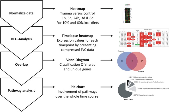
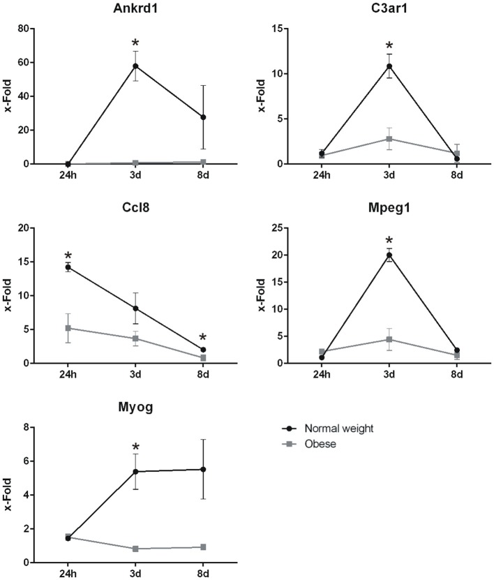
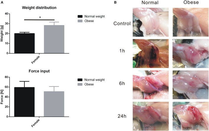
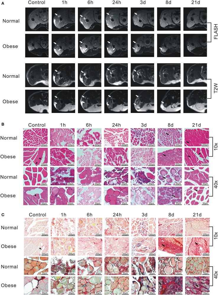
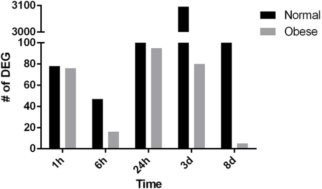
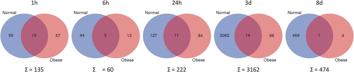
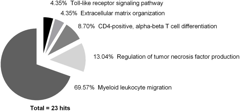
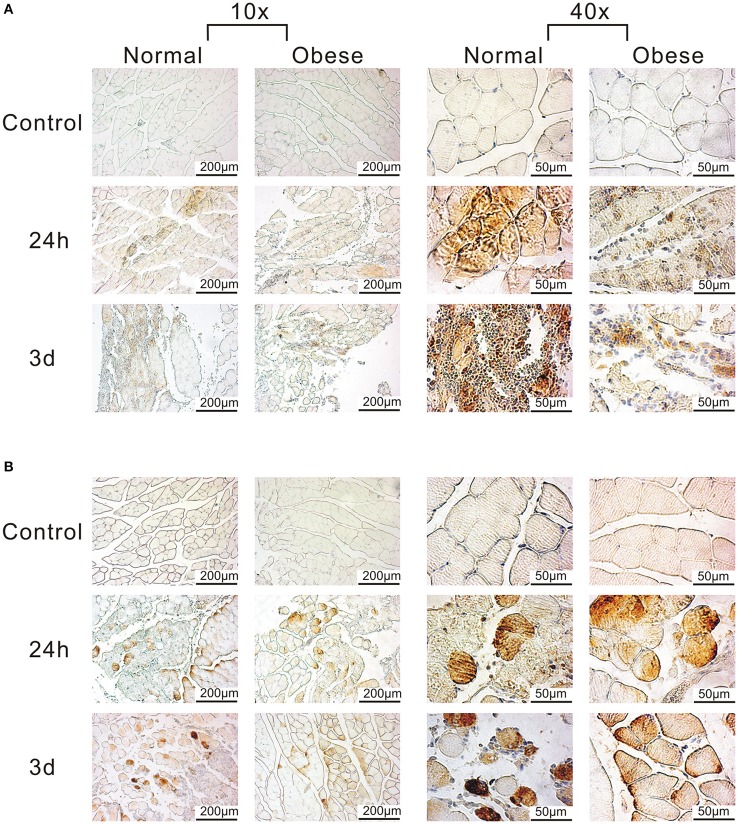
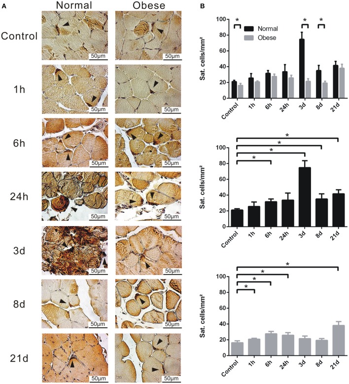
Injury to skeletal muscle affects millions of people worldwide. The underlying regenerative process however, is a very complex mechanism, time-wise highly coordinated, and subdivided in an initial inflammatory, a regenerative and a remodeling phase. Muscle regeneration can be impaired by several factors, among them diet-induced obesity (DIO). In order to evaluate if obesity negatively affects healing processes after trauma, we utilized a blunt injury approach to damage the muscle on the left hind limb of obese and normal weight C57BL/6J without showing any significant differences in force input between normal weight and obese mice. Magnetic resonance imaging (MRI) of the injury and regeneration process revealed edema formation and hemorrhage exudate in muscle tissue of normal weight and obese mice. In addition, morphological analysis of physiological changes revealed tissue necrosis, immune cell infiltration, extracellular matrix (ECM) remodeling, and fibrosis formation in the damaged muscle tissue. Regeneration was delayed in muscles of obese mice, with a higher incidence of fibrosis formation due to hampered expression levels of genes involved in ECM organization. Furthermore, a detailed molecular fingerprint in different stages of muscle regeneration underlined a delay or even lack of a regenerative response to injury in obese mice. A time-lapse heatmap determined 81 differentially expressed genes (DEG) with at least three hits in our model at all-time points, suggesting key candidates with a high impact on muscle regeneration. Pathway analysis of the DEG revealed five pathways with a high confidence level: myeloid leukocyte migration, regulation of tumor necrosis factor production, CD4-positive, alpha-beta T cell differentiation, ECM organization, and toll-like receptor (TLR) signaling. Moreover, changes in complement-, Wnt-, and satellite cell-related genes were found to be impaired in obese animals after trauma. Furthermore, histological satellite cell evaluation showed lower satellite cell numbers in the obese model upon injury. , and expression levels were also verified by qPCR. In summary, increased fibrosis formation, the reduction of satellite cells as well as specific changes in gene expression and signaling pathways could explain the delay of tissue regeneration in obese mice post trauma.
骨骼肌损伤影响着全球数百万人。然而,其潜在的再生过程是一个非常复杂的机制,在时间上高度协调,并分为初始炎症阶段、再生阶段和重塑阶段。肌肉再生会受到多种因素的影响,其中包括饮食诱导的肥胖(DIO)。为了评估肥胖是否会对创伤后的愈合过程产生负面影响,我们采用钝性损伤方法损伤肥胖和正常体重的C57BL/6J小鼠的左后肢肌肉,且正常体重和肥胖小鼠之间在力输入方面没有显示出任何显著差异。对损伤和再生过程进行磁共振成像(MRI)显示,正常体重和肥胖小鼠的肌肉组织中均出现水肿形成和出血渗出。此外,对生理变化的形态学分析显示,受损肌肉组织中存在组织坏死、免疫细胞浸润、细胞外基质(ECM)重塑和纤维化形成。肥胖小鼠的肌肉再生延迟,由于参与ECM组织的基因表达水平受阻,纤维化形成的发生率更高。此外,肌肉再生不同阶段的详细分子指纹图谱强调了肥胖小鼠对损伤的再生反应延迟甚至缺乏。时间推移热图确定了81个差异表达基因(DEG),在我们的模型中所有时间点至少有三次命中,表明这些关键候选基因对肌肉再生有很大影响。对DEG的通路分析揭示了五个具有高置信度的通路:髓样白细胞迁移、肿瘤坏死因子产生的调节、CD4阳性α-βT细胞分化、ECM组织和Toll样受体(TLR)信号传导。此外,发现肥胖动物创伤后补体、Wnt和卫星细胞相关基因的变化受损。此外,组织学卫星细胞评估显示,肥胖模型损伤后卫星细胞数量减少。 ,并且 表达水平也通过qPCR进行了验证。总之,纤维化形成增加、卫星细胞减少以及基因表达和信号通路的特定变化可以解释肥胖小鼠创伤后组织再生的延迟。