Department of Psychiatry and Behavioral Sciences, Johns Hopkins University School of Medicine, Baltimore, MD 21205, USA.
Center for Neurodevelopmental and Imaging Research, Kennedy Krieger Institute, Baltimore, MD 21205, USA; Department of Neurology, Johns Hopkins University School of Medicine, Baltimore, MD 21205, USA.
Neuroimage. 2022 Oct 15;260:119434. doi: 10.1016/j.neuroimage.2022.119434. Epub 2022 Jul 2.
Classic psychedelics, such as psilocybin and LSD, and other serotonin 2A receptor (5-HT) agonists evoke acute alterations in perception and cognition. Altered thalamocortical connectivity has been hypothesized to underlie these effects, which is supported by some functional MRI (fMRI) studies. These studies have treated the thalamus as a unitary structure, despite known differential 5-HT expression and functional specificity of different intrathalamic nuclei. Independent Component Analysis (ICA) has been previously used to identify reliable group-level functional subdivisions of the thalamus from resting-state fMRI (rsfMRI) data. We build on these efforts with a novel data-maximizing ICA-based approach to examine psilocybin-induced changes in intrathalamic functional organization and thalamocortical connectivity in individual participants.
Baseline rsfMRI data (n=38) from healthy individuals with a long-term meditation practice was utilized to generate a statistical template of thalamic functional subdivisions. This template was then applied in a novel ICA-based analysis of the acute effects of psilocybin on intra- and extra-thalamic functional organization and connectivity in follow-up scans from a subset of the same individuals (n=18). We examined correlations with subjective reports of drug effect and compared with a previously reported analytic approach (treating the thalamus as a single functional unit).
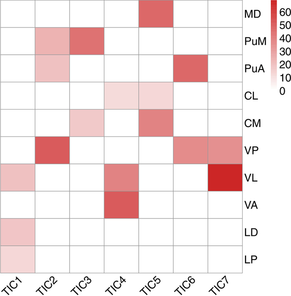
Several intrathalamic components showed significant psilocybin-induced alterations in spatial organization, with effects of psilocybin largely localized to the mediodorsal and pulvinar nuclei. The magnitude of changes in individual participants correlated with reported subjective effects. These components demonstrated predominant decreases in thalamocortical connectivity, largely with visual and default mode networks. Analysis in which the thalamus is treated as a singular unitary structure showed an overall numerical increase in thalamocortical connectivity, consistent with previous literature using this approach, but this increase did not reach statistical significance.
We utilized a novel analytic approach to discover psilocybin-induced changes in intra- and extra-thalamic functional organization and connectivity of intrathalamic nuclei and cortical networks known to express the 5-HT. These changes were not observed using whole-thalamus analyses, suggesting that psilocybin may cause widespread but modest increases in thalamocortical connectivity that are offset by strong focal decreases in functionally relevant intrathalamic nuclei.
经典迷幻剂,如裸盖菇素和 LSD,以及其他 5-羟色胺 2A 受体(5-HT)激动剂,会引起感知和认知的急性改变。改变丘脑皮质连接被认为是这些效应的基础,这得到了一些功能磁共振成像(fMRI)研究的支持。这些研究将丘脑视为一个单一的结构,尽管已知不同的丘脑核内有不同的 5-HT 表达和功能特异性。独立成分分析(ICA)之前已被用于从静息状态 fMRI(rsfMRI)数据中识别出丘脑的可靠的组水平功能细分。我们在这些努力的基础上,采用了一种新的基于数据最大化的 ICA 方法,以检查单个参与者中裸盖菇素诱导的丘脑内功能组织和丘脑皮质连接的变化。
利用有长期冥想练习的健康个体的基线 rsfMRI 数据(n=38)生成丘脑功能细分的统计模板。然后,在同一组个体的后续扫描中,使用一种新的基于 ICA 的分析方法,对裸盖菇素对内外丘脑功能组织和连接的急性影响进行分析(n=18)。我们检查了与药物效应的主观报告的相关性,并与以前的分析方法(将丘脑视为一个单一的功能单元)进行了比较。
几个丘脑内成分显示出明显的裸盖菇素诱导的空间组织变化,其作用主要局限于中背侧和豆状核。个体参与者变化的幅度与报告的主观效应相关。这些成分显示出丘脑皮质连接的显著减少,主要与视觉和默认模式网络有关。在将丘脑视为单一整体结构的分析中,显示出丘脑皮质连接的总体数值增加,与使用该方法的以前文献一致,但这种增加没有达到统计学意义。
我们利用一种新的分析方法,发现了裸盖菇素诱导的丘脑内核和皮质网络的内外功能组织和连接的变化,这些核和网络已知表达 5-HT。使用全丘脑分析没有观察到这些变化,这表明裸盖菇素可能导致广泛但适度的丘脑皮质连接增加,而功能相关的丘脑内核则强烈减少。