Yuan Rui, Di Xin, Taylor Paul A, Gohel Suril, Tsai Yuan-Hsiung, Biswal Bharat B
Department of Biomedical Engineering, New Jersey Institute of Technology, University Heights, Newark, NJ, 07102, USA.
Department of Electrical Engineering, New Jersey Institute of Technology, University Heights, Newark, NJ, 07102, USA.
Brain Struct Funct. 2016 May;221(4):1971-84. doi: 10.1007/s00429-015-1018-7. Epub 2015 Apr 30.
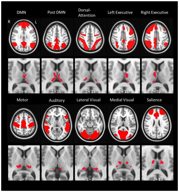
Various studies have indicated that the thalamus is involved in controlling both cortico-cortical information flow and cortical communication with the rest of the brain. Detailed anatomy and functional connectivity patterns of the thalamocortical system are essential to understanding the cortical organization and pathophysiology of a wide range of thalamus-related neurological and neuropsychiatric diseases. The current study used resting-state fMRI to investigate the topography of the human thalamocortical system from a functional perspective. The thalamus-related cortical networks were identified by performing independent component analysis on voxel-based thalamic functional connectivity maps across a large group of subjects. The resulting functional brain networks were very similar to well-established resting-state network maps. Using these brain network components in a spatial regression model with each thalamic voxel's functional connectivity map, we localized the thalamic subdivisions related to each brain network. For instance, the medial dorsal nucleus was shown to be associated with the default mode, the bilateral executive, the medial visual networks; and the pulvinar nucleus was involved in both the dorsal attention and the visual networks. These results revealed that a single nucleus may have functional connections with multiple cortical regions or even multiple functional networks, and may be potentially related to the function of mediation or modulation of multiple cortical networks. This observed organization of thalamocortical system provided a reference for studying the functions of thalamic sub-regions. The importance of intrinsic connectivity-based mapping of the thalamocortical relationship is discussed, as well as the applicability of the approach for future studies.
多项研究表明,丘脑参与控制皮质-皮质信息流以及皮质与大脑其他部分的通信。丘脑皮质系统的详细解剖结构和功能连接模式对于理解广泛的丘脑相关神经和神经精神疾病的皮质组织及病理生理学至关重要。当前研究使用静息态功能磁共振成像从功能角度研究人类丘脑皮质系统的拓扑结构。通过对一大组受试者基于体素的丘脑功能连接图进行独立成分分析,识别出与丘脑相关的皮质网络。所得的功能性脑网络与已确立的静息态网络图谱非常相似。在空间回归模型中,将这些脑网络成分与每个丘脑体素的功能连接图相结合,我们确定了与每个脑网络相关的丘脑亚区。例如,内侧背核被证明与默认模式、双侧执行、内侧视觉网络相关;枕核则参与背侧注意和视觉网络。这些结果表明,单个核可能与多个皮质区域甚至多个功能网络存在功能连接,并且可能潜在地与多个皮质网络的介导或调节功能相关。这种观察到的丘脑皮质系统组织为研究丘脑亚区的功能提供了参考。文中还讨论了基于内在连接性绘制丘脑皮质关系图的重要性,以及该方法在未来研究中的适用性。