Department of Geriatrics, The First Affiliated Hospital of Soochow University, Suzhou, Jiangsu Province, China.
Key Laboratory for Aging & Disease, Nanjing Medical University, Nanjing, Jiangsu Province, China.
Dis Markers. 2022 Jun 27;2022:1919064. doi: 10.1155/2022/1919064. eCollection 2022.
Cyclin-dependent kinase-5 (CDK5) is a key kinase involved in brain development and function and recently found to be involved in neuronal and astroglial apoptosis, neural stem/progenitor cell stemness, mitochondrial fission, and synaptic transmission. But the specific mechanism of CDK5-mediated anti-inflammatory remains unclear in ICH. The aim of the present study was to explore the role of CDK5 in mediating microglia activity through activated DRP1 phosphorylation in a rat ICH model.
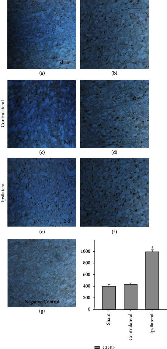
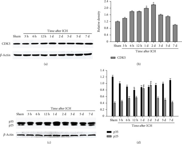
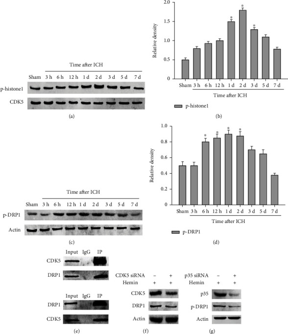
We measured behavioral change after ICH; detected the expression of CDK5 in the rat brain using immunohistochemistry; and measured the protein levels of CDK5, p35, p25, p-histone H1, and p-DRP1 using Western blot analysis. Coimmunoprecipitation analysis indicated interaction of CDK5 and DRP1. Tumor necrosis factor-, interleukin- (IL-) 1, and IL-6 levels were measured using enzyme-linked immunosorbent assay (ELISA).
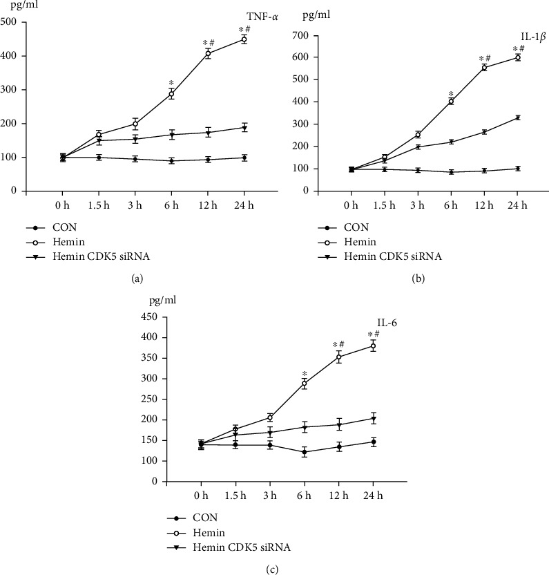
After ICH, CDK5 protein level and kinase activity increased. Western blot data showed that CDK5 expression increased from 6 h and peaked at 2 d after ICH ( < 0.05), and the expression of p35 was lowest at 12 h, while the expression of p25 peaked at 2 d after ICH. Besides, p-DRP1 expression change follows with CDK5 kinase activity change. Coimmunoprecipitation showed that interaction between CDK5 and DRP1 certainly exists in microglia. Then, knockdown CDK5 or p35 expression by siRNA reduced the expression level of p-DRP1. ELISA data showed that the protein levels of proinflammatory mediators, such as TNF-, IL-1, and IL-6, were decreased by knockdown of CDK5.
CDK5 may regulate DRP1 by direct phosphorylation in microglia and further induce microglia secreting proinflammation factor.
周期蛋白依赖性激酶-5(CDK5)是一种参与脑发育和功能的关键激酶,最近发现其参与神经元和星形胶质细胞凋亡、神经干细胞/祖细胞干性、线粒体裂变和突触传递。但 CDK5 介导抗炎的具体机制在 ICH 中尚不清楚。本研究旨在通过在大鼠 ICH 模型中研究 CDK5 通过激活 DRP1 磷酸化调节小胶质细胞活性的作用来探索其作用机制。
我们测量了 ICH 后的行为变化;通过免疫组织化学检测大鼠脑内 CDK5 的表达;并通过 Western blot 分析检测 CDK5、p35、p25、p-H1 组蛋白和 p-DRP1 的蛋白水平。免疫共沉淀分析表明 CDK5 和 DRP1 相互作用。使用酶联免疫吸附测定(ELISA)测量肿瘤坏死因子-α、白细胞介素-(IL-)1 和 IL-6 的水平。
ICH 后,CDK5 蛋白水平和激酶活性增加。Western blot 数据显示,CDK5 表达从 6 小时开始增加,并在 ICH 后 2 天达到峰值(<0.05),p35 的表达在 12 小时最低,而 p25 的表达在 ICH 后 2 天达到峰值。此外,p-DRP1 的表达变化与 CDK5 激酶活性变化一致。免疫共沉淀显示,CDK5 和 DRP1 之间确实存在相互作用。然后,通过 siRNA 敲低 CDK5 或 p35 表达降低了 p-DRP1 的表达水平。ELISA 数据显示,通过敲低 CDK5,促炎介质如 TNF-α、IL-1 和 IL-6 的蛋白水平降低。
CDK5 可能通过直接磷酸化小胶质细胞中的 DRP1 来调节 DRP1,进而诱导小胶质细胞分泌促炎因子。