Instituto de Estudios Inmunológicos y Fisiopatológicos (IIFP), UNLP, CONICET, Asociado a CIC PBA, Facultad de Ciencias Exactas, Departamento de Ciencias Biológicas, La Plata, Argentina.
Servicio de Gastroenterología, Hospital de Niños Sor María Ludovica, La Plata, Argentina.
Front Immunol. 2022 Jun 21;13:909896. doi: 10.3389/fimmu.2022.909896. eCollection 2022.
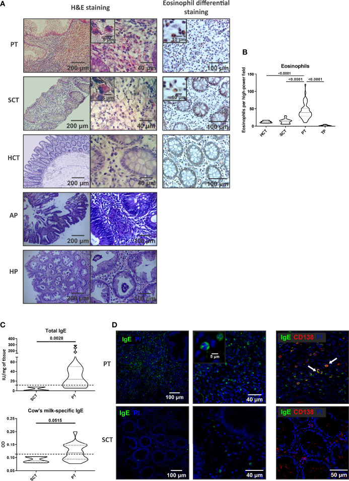
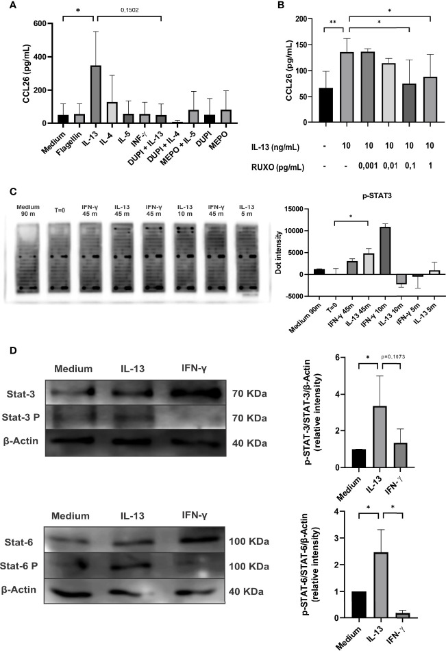
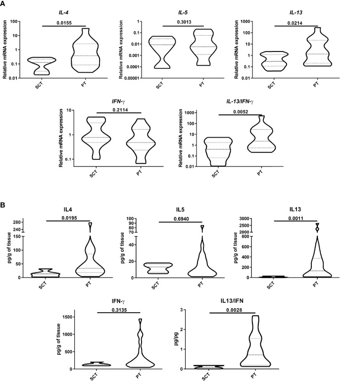
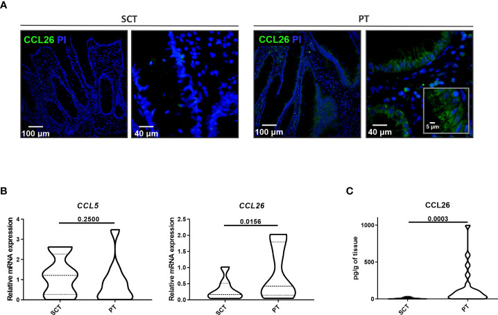
Several inflammatory processes of the bowel are characterized by an accumulation of eosinophils at inflammation sites. The mechanisms that govern mucosal infiltration with eosinophils are not fully understood. In this work, we studied the colorectal polyp-confined tissue containing eosinophils and we hypothesized that intestinal epithelial cells are the cell source of eotaxin-3 or CCL26, a potent chemoattractant for eosinophils. We analyzed colorectal polyps (n=50) from pediatric patients with rectal bleeding by H&E staining and eosin staining, and different pro-inflammatory cytokines were assessed by RT-qPCR and ELISA. IgE and CCL26 were investigated by RT-qPCR, ELISA and confocal microscopy. Finally, the intracellular signaling pathway that mediates the CCL26 production was analyzed using a kinase array and immunoblotting in human intestinal Caco-2 cell line. We found a dense cell agglomeration within the polyps, with a significantly higher frequency of eosinophils than in control adjacent tissue. IL-4 and IL-13 were significantly up-regulated in polyps and CCL26 was elevated in the epithelial compartment. Experiments with Caco-2 cells showed that the type-2 cytokine IL-13 increased STAT3 and STAT6 phosphorylation and eotaxin-3 secretion. The addition of the blocking antibody Dupilumab or the inhibitor Ruxolitinib to the cytokine-stimulated Caco-2 cells diminished the CCL26 secretion to basal levels in a dose-dependent manner. In conclusion, our findings demonstrate a high frequency of eosinophils, and elevated levels of type-2 cytokines and eotaxin-3 in the inflammatory stroma of colorectal polyps from pediatric patients. Polyp epithelial cells showed to be the main cell source of CCL26, and IL-13 was the main trigger of this chemokine through the activation of the STAT3/STAT6/JAK1-2 pathway. We suggest that the epithelial compartment actively participates in the recruitment of eosinophils to the colonic polyp-confined inflammatory environment.
几种肠道炎症过程的特点是炎症部位嗜酸性粒细胞的积累。控制嗜酸性粒细胞黏膜浸润的机制尚未完全阐明。在这项工作中,我们研究了含有嗜酸性粒细胞的结肠直肠息肉局限组织,并假设肠上皮细胞是嗜酸性粒细胞趋化因子 eotaxin-3 或 CCL26 的细胞来源。我们通过 H&E 染色和嗜酸性粒细胞染色分析了来自直肠出血的儿科患者的结肠直肠息肉(n=50),并通过 RT-qPCR 和 ELISA 评估了不同的促炎细胞因子。通过 RT-qPCR、ELISA 和共聚焦显微镜研究了 IgE 和 CCL26。最后,使用激酶阵列和免疫印迹分析了在人肠 Caco-2 细胞系中介导 CCL26 产生的细胞内信号通路。我们发现息肉内有密集的细胞聚集,嗜酸性粒细胞的频率明显高于相邻对照组织。IL-4 和 IL-13 在息肉中显著上调,上皮细胞区 CCL26 升高。用 Caco-2 细胞进行的实验表明,2 型细胞因子 IL-13 增加了 STAT3 和 STAT6 的磷酸化和 eotaxin-3 的分泌。向细胞因子刺激的 Caco-2 细胞中添加阻断抗体 Dupilumab 或抑制剂 Ruxolitinib 以剂量依赖性方式将 CCL26 分泌减少到基础水平。总之,我们的研究结果表明,在儿科患者的结肠直肠息肉炎症基质中,嗜酸性粒细胞、2 型细胞因子和 eotaxin-3 的水平均较高。息肉上皮细胞显示为 CCL26 的主要细胞来源,IL-13 通过激活 STAT3/STAT6/JAK1-2 通路成为这种趋化因子的主要触发因素。我们认为上皮细胞区积极参与招募嗜酸性粒细胞进入结肠息肉局限的炎症环境。