Lysenko Anna, Razumova Alexandra, Yaremenko Andrey, Ivanov Vladimir, Strelkov Sergey, Krivtsov Anton
Department of Dental Surgery and Maxillofacial Surgery, First Pavlov State Medical University, Saint-Petersburg, Russia.
Higher School of Theoretical Mechanics, Peter the Great Saint Petersburg Polytechnic University, Saint-Petersburg, Russia.
Imaging Sci Dent. 2022 Jun;52(2):225-230. doi: 10.5624/isd.20210256. Epub 2022 Mar 15.
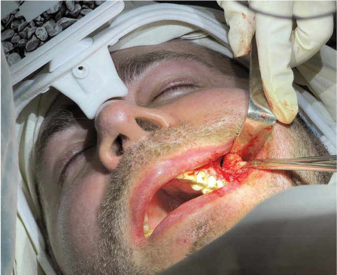
This report presents the first known use of a rigid endoscope with augmented reality technology for the removal of an odontogenic cyst that penetrated the maxillary sinus and illustrates its practical use in a patient.
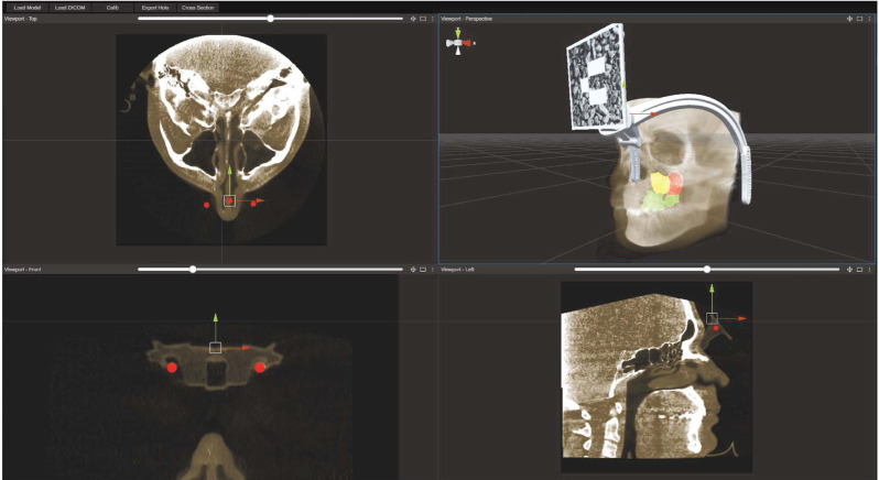
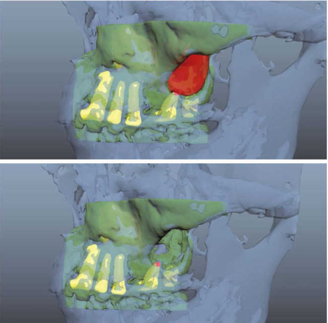
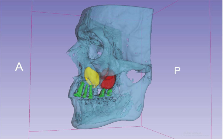
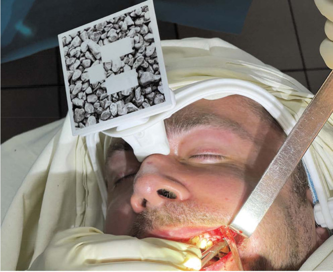
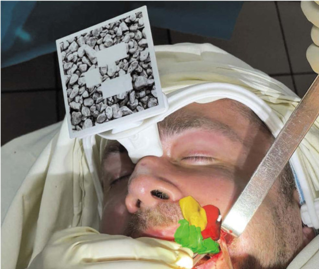
In the preoperative period, cone-beam computed tomography was performed in a specially designed marker holder frame, and the contours of the cyst and the nearest anatomical formations were segmented in the 3D Slicer program. During the operation, a marker was installed on the patient's head, as well as on the tip of the endoscope, which made it possible to visualize the mass and the movement of the endoscope. The surgical intervention was performed with the support of augmented reality in HoloLens glasses (Microsoft Corporation, Redmond, WA, USA).
The use of this technology improved the accuracy of surgical manipulations, reduced operational risks, and shortened the time of surgery and the rehabilitation period.
With the help of modern technologies, a navigation system was created that helped to track the position of the endoscope in mixed reality in real time, as well as to fully visualize anatomical formations.
本报告介绍了首次使用带有增强现实技术的硬质内窥镜切除穿透上颌窦的牙源性囊肿,并阐述了其在一名患者中的实际应用。
术前,在专门设计的标记支架框架中进行锥形束计算机断层扫描,并在3D Slicer程序中分割囊肿和最接近的解剖结构轮廓。手术过程中,在患者头部以及内窥镜尖端安装标记,这使得能够可视化肿块和内窥镜的移动。手术干预在HoloLens眼镜(美国华盛顿州雷德蒙德市微软公司)的增强现实支持下进行。
该技术的使用提高了手术操作的准确性,降低了手术风险,缩短了手术时间和康复期。
借助现代技术,创建了一个导航系统,有助于实时跟踪内窥镜在混合现实中的位置,并能完全可视化解剖结构。