• 文献检索
  • 文档翻译
  • 深度研究
  • 学术资讯
  • Suppr Zotero 插件Zotero 插件
  • 邀请有礼
  • 套餐&价格
  • 历史记录
应用&插件
Suppr Zotero 插件Zotero 插件浏览器插件Mac 客户端Windows 客户端微信小程序
定价
高级版会员购买积分包购买API积分包
服务
文献检索文档翻译深度研究API 文档MCP 服务
关于我们
关于 Suppr公司介绍联系我们用户协议隐私条款
关注我们

Suppr 超能文献

核心技术专利:CN118964589B侵权必究
粤ICP备2023148730 号-1Suppr @ 2026

文献检索

告别复杂PubMed语法,用中文像聊天一样搜索,搜遍4000万医学文献。AI智能推荐,让科研检索更轻松。

立即免费搜索

文件翻译

保留排版,准确专业,支持PDF/Word/PPT等文件格式,支持 12+语言互译。

免费翻译文档

深度研究

AI帮你快速写综述,25分钟生成高质量综述,智能提取关键信息,辅助科研写作。

立即免费体验

高灵敏度器官靶向 PET 相机的评估。

Evaluation of a High-Sensitivity Organ-Targeted PET Camera.

机构信息

Department of Physics, Lakehead University, Thunder Bay, ON P7B 5E1, Canada.

Radialis Medical, Thunder Bay, ON P7A 7T1, Canada.

出版信息

Sensors (Basel). 2022 Jun 21;22(13):4678. doi: 10.3390/s22134678.

DOI:10.3390/s22134678
PMID:35808181
原文链接:https://pmc.ncbi.nlm.nih.gov/articles/PMC9269056/
Abstract

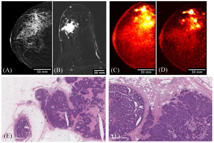
The aim of this study is to evaluate the performance of the Radialis organ-targeted positron emission tomography (PET) Camera with standardized tests and through assessment of clinical-imaging results. Sensitivity, count-rate performance, and spatial resolution were evaluated according to the National Electrical Manufacturers Association (NEMA) NU-4 standards, with necessary modifications to accommodate the planar detector design. The detectability of small objects was shown with micro hotspot phantom images. The clinical performance of the camera was also demonstrated through breast cancer images acquired with varying injected doses of 2-[fluorine-18]-fluoro-2-deoxy-D-glucose (F-FDG) and qualitatively compared with sample digital full-field mammography, magnetic resonance imaging (MRI), and whole-body (WB) PET images. Micro hotspot phantom sources were visualized down to 1.35 mm-diameter rods. Spatial resolution was calculated to be 2.3 ± 0.1 mm for the in-plane resolution and 6.8 ± 0.1 mm for the cross-plane resolution using maximum likelihood expectation maximization (MLEM) reconstruction. The system peak noise equivalent count rate was 17.8 kcps at a F-FDG concentration of 10.5 kBq/mL. System scatter fraction was 24%. The overall efficiency at the peak noise equivalent count rate was 5400 cps/MBq. The maximum axial sensitivity achieved was 3.5%, with an average system sensitivity of 2.4%. Selected results from clinical trials demonstrate capability of imaging lesions at the chest wall and identifying false-negative X-ray findings and false-positive MRI findings, even at up to a 10-fold dose reduction in comparison with standard F-FDG doses (i.e., at 37 MBq or 1 mCi). The evaluation of the organ-targeted Radialis PET Camera indicates that it is a promising technology for high-image-quality, low-dose PET imaging. High-efficiency radiotracer detection also opens an opportunity to reduce administered doses of radiopharmaceuticals and, therefore, patient exposure to radiation.

摘要

本研究旨在通过标准化测试和临床成像结果评估,评估 Radialis 器官靶向正电子发射断层扫描(PET)相机的性能。根据美国电器制造商协会(NEMA)NU-4 标准,对灵敏度、计数率性能和空间分辨率进行了评估,并对平面探测器设计进行了必要的修改。通过微热点体模图像显示了对小物体的探测能力。还通过不同注射剂量的 2-[氟-18]-氟-2-脱氧-D-葡萄糖(F-FDG)获得的乳腺癌图像来展示相机的临床性能,并与样本数字全视野乳房 X 线摄影、磁共振成像(MRI)和全身(WB)PET 图像进行定性比较。可以观察到直径为 1.35 毫米的微热点体模源。使用最大似然期望最大化(MLEM)重建,计算得到平面内分辨率为 2.3 ± 0.1 毫米,交叉平面分辨率为 6.8 ± 0.1 毫米。在 F-FDG 浓度为 10.5 kBq/mL 时,系统峰值噪声等效计数率为 17.8 kcps。系统散射分数为 24%。在峰值噪声等效计数率下,总效率为 5400 cps/MBq。达到的最大轴向灵敏度为 3.5%,平均系统灵敏度为 2.4%。临床试验的部分结果表明,该相机能够对胸壁病变进行成像,并识别 X 线检查的假阴性和 MRI 检查的假阳性结果,即使与标准 F-FDG 剂量相比,剂量降低 10 倍(即 37 MBq 或 1 mCi)也是如此。对器官靶向 Radialis PET 相机的评估表明,它是一种用于高质量、低剂量 PET 成像的有前途的技术。高效的放射性示踪剂检测还为减少放射性药物的给药剂量提供了机会,从而减少了患者的辐射暴露。

https://cdn.ncbi.nlm.nih.gov/pmc/blobs/5a7a/9269056/caa63fcd9ec4/sensors-22-04678-g014.jpg
https://cdn.ncbi.nlm.nih.gov/pmc/blobs/5a7a/9269056/613a9d239e3e/sensors-22-04678-g001.jpg
https://cdn.ncbi.nlm.nih.gov/pmc/blobs/5a7a/9269056/31fa03241811/sensors-22-04678-g002.jpg
https://cdn.ncbi.nlm.nih.gov/pmc/blobs/5a7a/9269056/ba34c62cbab6/sensors-22-04678-g003.jpg
https://cdn.ncbi.nlm.nih.gov/pmc/blobs/5a7a/9269056/663bc3540a1a/sensors-22-04678-g004.jpg
https://cdn.ncbi.nlm.nih.gov/pmc/blobs/5a7a/9269056/ebff612bf9cf/sensors-22-04678-g005.jpg
https://cdn.ncbi.nlm.nih.gov/pmc/blobs/5a7a/9269056/a9d3df7222e4/sensors-22-04678-g006.jpg
https://cdn.ncbi.nlm.nih.gov/pmc/blobs/5a7a/9269056/f37f04f3fa4e/sensors-22-04678-g007.jpg
https://cdn.ncbi.nlm.nih.gov/pmc/blobs/5a7a/9269056/8e86658677eb/sensors-22-04678-g008.jpg
https://cdn.ncbi.nlm.nih.gov/pmc/blobs/5a7a/9269056/d6906f10dc49/sensors-22-04678-g009.jpg
https://cdn.ncbi.nlm.nih.gov/pmc/blobs/5a7a/9269056/c7372c0007ce/sensors-22-04678-g010.jpg
https://cdn.ncbi.nlm.nih.gov/pmc/blobs/5a7a/9269056/ad7fa4d28b51/sensors-22-04678-g011.jpg
https://cdn.ncbi.nlm.nih.gov/pmc/blobs/5a7a/9269056/c4ad136f48b2/sensors-22-04678-g012.jpg
https://cdn.ncbi.nlm.nih.gov/pmc/blobs/5a7a/9269056/968563a1fb51/sensors-22-04678-g013.jpg
https://cdn.ncbi.nlm.nih.gov/pmc/blobs/5a7a/9269056/caa63fcd9ec4/sensors-22-04678-g014.jpg

相似文献

1
Evaluation of a High-Sensitivity Organ-Targeted PET Camera.高灵敏度器官靶向 PET 相机的评估。
Sensors (Basel). 2022 Jun 21;22(13):4678. doi: 10.3390/s22134678.
2
Technical Note: Performance evaluation of a small-animal PET/CT system based on NEMA NU 4-2008 standards.技术说明:基于 NEMA NU 4-2008 标准的小动物 PET/CT 系统性能评估。
Med Phys. 2021 Sep;48(9):5272-5282. doi: 10.1002/mp.15088. Epub 2021 Jul 26.
3
An extended bore length solid-state digital-BGO PET/CT system: design, preliminary experience, and performance characteristics.一种扩展孔径长度的固态数字 BGO PET/CT 系统:设计、初步经验和性能特点。
Eur J Nucl Med Mol Imaging. 2024 Mar;51(4):954-964. doi: 10.1007/s00259-023-06514-8. Epub 2023 Nov 28.
4
Performance evaluation of the inveon dedicated PET preclinical tomograph based on the NEMA NU-4 standards.基于NEMA NU-4标准对inveon专用PET临床前断层扫描仪的性能评估。
J Nucl Med. 2009 Mar;50(3):401-8. doi: 10.2967/jnumed.108.056374. Epub 2009 Feb 17.
5
Performance characteristics of a new 3-dimensional continuous-emission and spiral-transmission high-sensitivity and high-resolution PET camera evaluated with the NEMA NU 2-2001 standard.采用NEMA NU 2-2001标准评估的新型三维连续发射和螺旋传输高灵敏度高分辨率PET相机的性能特征。
J Nucl Med. 2006 Jan;47(1):83-90.
6
Performance Characteristics of a New-Generation Digital Bismuth Germanium Oxide PET/CT System, Omni Legend 32, According to NEMA NU 2-2018 Standards.新一代数字化锗铋氧化物 PET/CT 系统(奥米龙 32 型)的性能特征,符合 NEMA NU 2-2018 标准。
J Nucl Med. 2023 Dec 1;64(12):1990-1997. doi: 10.2967/jnumed.123.266140.
7
Performance Characteristics of the NeuroEXPLORER, a Next-Generation Human Brain PET/CT Imager.新一代人脑正电子发射断层显像/计算机断层扫描(PET/CT)成像仪NeuroEXPLORER的性能特征
J Nucl Med. 2024 Aug 1;65(8):1320-1326. doi: 10.2967/jnumed.124.267767.
8
More advantages in detecting bone and soft tissue metastases from prostate cancer using F-PSMA PET/CT.使用F-PSMA PET/CT检测前列腺癌骨和软组织转移方面有更多优势。
Hell J Nucl Med. 2019 Jan-Apr;22(1):6-9. doi: 10.1967/s002449910952. Epub 2019 Mar 7.
9
NEMA NU-04-based performance characteristics of the LabPET-8™ small animal PET scanner.基于 NEMA NU-04 的 LabPET-8™ 小动物 PET 扫描仪性能特征。
Phys Med Biol. 2011 Oct 21;56(20):6649-64. doi: 10.1088/0031-9155/56/20/009. Epub 2011 Sep 23.
10
Monte Carlo evaluation of hypothetical long axial field-of-view PET scanner using GE Discovery MI PET front-end architecture.使用 GE Discovery MI PET 前端架构对假设的长轴向视野 PET 扫描仪进行蒙特卡罗评估。
Med Phys. 2022 Feb;49(2):1139-1152. doi: 10.1002/mp.15422. Epub 2022 Jan 10.

引用本文的文献

1
Translating lifestyle interventions for optimal brain health in Africa.将生活方式干预措施应用于非洲以实现最佳脑健康状况的转化。
Nat Rev Neurol. 2025 Jul 11. doi: 10.1038/s41582-025-01104-8.
2
Multi-angle acquisition and 3D composite reconstruction for organ-targeted PET using planar detectors.使用平面探测器进行器官靶向PET的多角度采集和三维复合重建。
Med Phys. 2025 Apr;52(4):2507-2519. doi: 10.1002/mp.17606. Epub 2024 Dec 27.
3
Case report: Possible role of low-dose PEM for avoiding unneeded procedures associated with false-positive or equivocal breast MRI results.

本文引用的文献

1
Downstream Mammary and Extramammary Cascade Services and Spending Following Screening Breast Magnetic Resonance Imaging vs Mammography Among Commercially Insured Women.商业保险女性接受筛查性乳腺磁共振成像与乳腺 X 线摄影后下游乳腺和乳腺外级联服务及支出。
JAMA Netw Open. 2022 Apr 1;5(4):e227234. doi: 10.1001/jamanetworkopen.2022.7234.
2
Deep learning-assisted PET imaging achieves fast scan/low-dose examination.深度学习辅助的正电子发射断层显像(PET)成像可实现快速扫描/低剂量检查。
EJNMMI Phys. 2022 Feb 4;9(1):7. doi: 10.1186/s40658-022-00431-9.
3
Contemporary issues in radiation protection in medical imaging: introductory editorial.
病例报告:低剂量钆双胺增强磁共振成像在避免与乳腺磁共振成像假阳性或不确定结果相关的不必要检查中的潜在作用
Front Oncol. 2024 Jul 18;14:1405404. doi: 10.3389/fonc.2024.1405404. eCollection 2024.
4
Image quality evaluation for a clinical organ-targeted PET camera.临床器官靶向正电子发射断层显像(PET)相机的图像质量评估
Front Oncol. 2024 Mar 25;14:1268991. doi: 10.3389/fonc.2024.1268991. eCollection 2024.
5
Hyperpolarized Xenon-129 Chemical Exchange Saturation Transfer (HyperCEST) Molecular Imaging: Achievements and Future Challenges.氙-129 超极化化学交换饱和传递(HyperCEST)分子成像:成就与未来挑战。
Int J Mol Sci. 2024 Feb 5;25(3):1939. doi: 10.3390/ijms25031939.
6
Breast Cancer Detection Using a Low-Dose Positron Emission Digital Mammography System.使用低剂量正电子发射数字乳腺摄影系统进行乳腺癌检测。
Radiol Imaging Cancer. 2024 Mar;6(2):e230020. doi: 10.1148/rycan.230020.
7
The quest for multifunctional and dedicated PET instrumentation with irregular geometries.追求具有不规则几何形状的多功能、专用 PET 仪器。
Ann Nucl Med. 2024 Jan;38(1):31-70. doi: 10.1007/s12149-023-01881-6. Epub 2023 Nov 12.
8
A look at radiation detectors and their applications in medical imaging.探讨辐射探测器及其在医学成像中的应用。
Jpn J Radiol. 2024 Feb;42(2):145-157. doi: 10.1007/s11604-023-01486-z. Epub 2023 Sep 21.
医学成像中辐射防护的当代问题:引言社论
Br J Radiol. 2021 Oct;94(1126):bjr20219004. doi: 10.1259/bjr.20219004.
4
Low-count whole-body PET with deep learning in a multicenter and externally validated study.在一项多中心且经过外部验证的研究中,利用深度学习进行低计数全身正电子发射断层扫描。
NPJ Digit Med. 2021 Aug 23;4(1):127. doi: 10.1038/s41746-021-00497-2.
5
Performance Characteristics of the Biograph Vision Quadra PET/CT System with a Long Axial Field of View Using the NEMA NU 2-2018 Standard.使用NEMA NU 2-2018标准对具有长轴向视野的Biograph Vision Quadra PET/CT系统的性能特征进行研究。
J Nucl Med. 2022 Mar;63(3):476-484. doi: 10.2967/jnumed.121.261972. Epub 2021 Jul 22.
6
Deep learning-assisted ultra-fast/low-dose whole-body PET/CT imaging.深度学习辅助的超快速/低剂量全身 PET/CT 成像。
Eur J Nucl Med Mol Imaging. 2021 Jul;48(8):2405-2415. doi: 10.1007/s00259-020-05167-1. Epub 2021 Jan 25.
7
Evaluation of primary breast cancers using dedicated breast PET and whole-body PET.使用专用乳腺 PET 和全身 PET 对原发性乳腺癌进行评估。
Sci Rep. 2020 Dec 14;10(1):21930. doi: 10.1038/s41598-020-78865-3.
8
The Future of Nuclear Medicine, Molecular Imaging, and Theranostics.核医学、分子影像与诊疗一体化的未来
J Nucl Med. 2020 Dec;61(Suppl 2):263S-272S. doi: 10.2967/jnumed.120.254532.
9
State of the art in total body PET.全身正电子发射断层扫描(PET)的技术现状
EJNMMI Phys. 2020 May 25;7(1):35. doi: 10.1186/s40658-020-00290-2.
10
Gaussian position-weighted center of gravity algorithm for multiplexed readout.高斯位置加权重心算法用于多重读出。
Phys Med Biol. 2020 Aug 11;65(16):165003. doi: 10.1088/1361-6560/ab9357.