• 文献检索
  • 文档翻译
  • 深度研究
  • 学术资讯
  • Suppr Zotero 插件Zotero 插件
  • 邀请有礼
  • 套餐&价格
  • 历史记录
应用&插件
Suppr Zotero 插件Zotero 插件浏览器插件Mac 客户端Windows 客户端微信小程序
定价
高级版会员购买积分包购买API积分包
服务
文献检索文档翻译深度研究API 文档MCP 服务
关于我们
关于 Suppr公司介绍联系我们用户协议隐私条款
关注我们

Suppr 超能文献

核心技术专利:CN118964589B侵权必究
粤ICP备2023148730 号-1Suppr @ 2026

文献检索

告别复杂PubMed语法,用中文像聊天一样搜索,搜遍4000万医学文献。AI智能推荐,让科研检索更轻松。

立即免费搜索

文件翻译

保留排版,准确专业,支持PDF/Word/PPT等文件格式,支持 12+语言互译。

免费翻译文档

深度研究

AI帮你快速写综述,25分钟生成高质量综述,智能提取关键信息,辅助科研写作。

立即免费体验

全身正电子发射断层扫描(PET)的技术现状

State of the art in total body PET.

作者信息

Vandenberghe Stefaan, Moskal Pawel, Karp Joel S

机构信息

Department of Electronics and Information Systems, MEDISIP, Ghent University-IBiTech, De Pintelaan 185 block B, Ghent, B-9000, Belgium.

Institute of Physics, Jagiellonian University, Krakow, Poland.

出版信息

EJNMMI Phys. 2020 May 25;7(1):35. doi: 10.1186/s40658-020-00290-2.

DOI:10.1186/s40658-020-00290-2
PMID:32451783
原文链接:https://pmc.ncbi.nlm.nih.gov/articles/PMC7248164/
Abstract

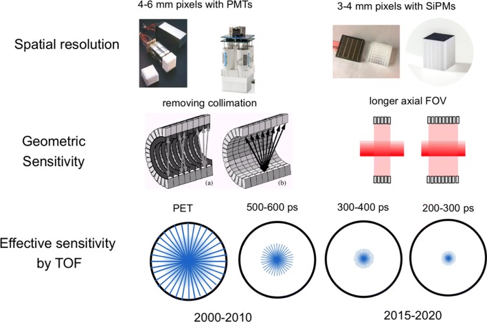
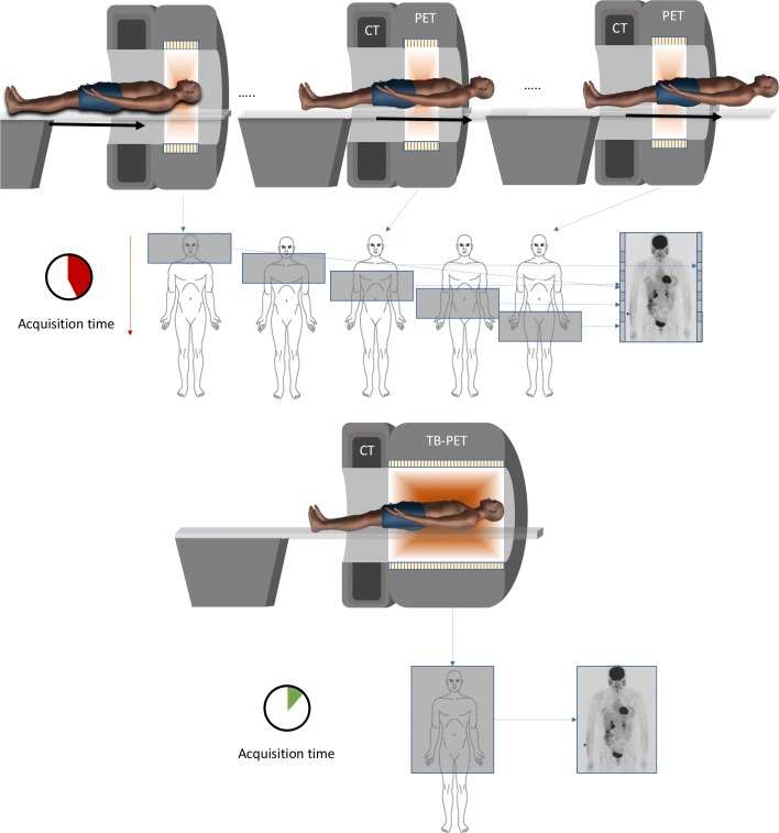
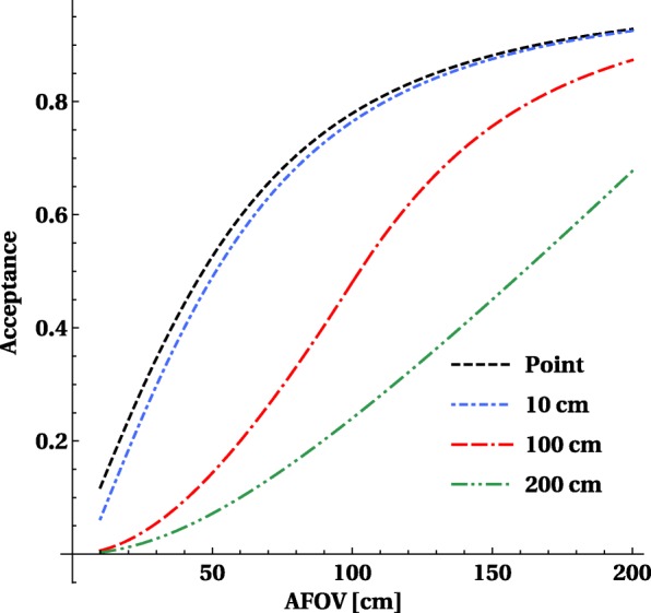
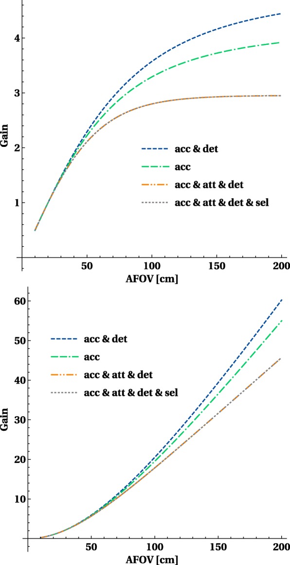
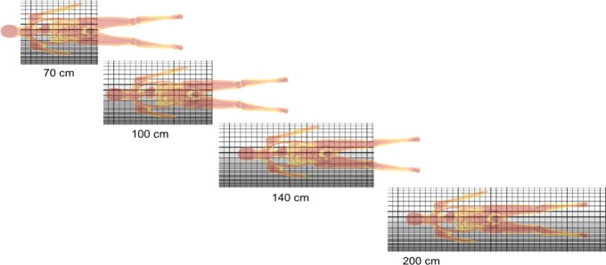
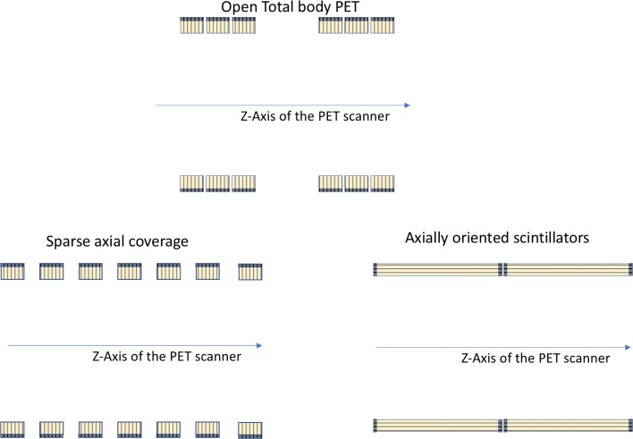
The idea of a very sensitive positron emission tomography (PET) system covering a large portion of the body of a patient already dates back to the early 1990s. In the period 2000-2010, only some prototypes with long axial field of view (FOV) have been built, which never resulted in systems used for clinical research. One of the reasons was the limitations in the available detector technology, which did not yet have sufficient energy resolution, timing resolution or countrate capabilities for fully exploiting the benefits of a long axial FOV design. PET was also not yet as widespread as it is today: the growth in oncology, which has become the major application of PET, appeared only after the introduction of PET-CT (early 2000).The detector technology used in most clinical PET systems today has a combination of good energy and timing resolution with higher countrate capabilities and has now been used since more than a decade to build time-of-flight (TOF) PET systems with fully 3D acquisitions. Based on this technology, one can construct total body PET systems and the remaining challenges (data handling, fast image reconstruction, detector cooling) are mostly related to engineering. The direct benefits of long axial FOV systems are mostly related to the higher sensitivity. For single organ imaging, the gain is close to the point source sensitivity which increases linearly with the axial length until it is limited by solid angle and attenuation of the body. The gains for single organ (compared to a fully 3D PET 20-cm axial FOV) are limited to a factor 3-4. But for long objects (like body scans), it increases quadratically with scanner length and factors of 10-40 × higher sensitivity are predicted for the long axial FOV scanner. This application of PET has seen a major increase (mostly in oncology) during the last 2 decades and is now the main type of study in a PET centre. As the technology is available and the full body concept also seems to match with existing applications, the old concept of a total body PET scanner is seeing a clear revival. Several research groups are working on this concept and after showing the potential via extensive simulations; construction of these systems has started about 2 years ago. In the first phase, two PET systems with long axial FOV suitable for large animal imaging were constructed to explore the potential in more experimental settings. Recently, the first completed total body PET systems for human use, a 70-cm-long system, called PennPET Explorer, and a 2-m-long system, called uExplorer, have become reality and first clinical studies have been shown. These results illustrate the large potential of this concept with regard to low-dose imaging, faster scanning, whole-body dynamic imaging and follow-up of tracers over longer periods. This large range of possible technical improvements seems to have the potential to change the current clinical routine and to expand the number of clinical applications of molecular imaging. The J-PET prototype is a prototype system with a long axial FOV built from axially arranged plastic scintillator strips.This paper gives an overview of the recent technical developments with regard to PET scanners with a long axial FOV covering at least the majority of the body (so called total body PET systems). After explaining the benefits and challenges of total body PET systems, the different total body PET system designs proposed for large animal and clinical imaging are described in detail. The axial length is one of the major factors determining the total cost of the system, but there are also other options in detector technology, design and processing for reducing the cost these systems. The limitations and advantages of different designs for research and clinical use are discussed taking into account potential applications and the increased cost of these systems.

摘要

早在20世纪90年代初,就已经有了关于构建一种能够覆盖患者身体大部分区域的高灵敏度正电子发射断层扫描(PET)系统的想法。在2000年至2010年期间,仅制造了一些具有长轴向视野(FOV)的原型机,但这些原型机从未发展成为用于临床研究的系统。其中一个原因是当时可用的探测器技术存在局限性,其能量分辨率、时间分辨率或计数率能力尚不足以充分利用长轴向FOV设计的优势。此外,PET在当时也不像现在这样普及:肿瘤学领域的发展,而肿瘤学如今已成为PET的主要应用领域,是在PET-CT推出后(2000年初)才出现的。如今大多数临床PET系统所使用的探测器技术,具备良好的能量和时间分辨率,同时计数率能力更高,并且在过去十多年里一直用于构建具有全三维采集功能的飞行时间(TOF)PET系统。基于这项技术,可以构建全身PET系统,而剩下的挑战(数据处理、快速图像重建、探测器冷却)大多与工程技术相关。长轴向FOV系统的直接优势主要与更高的灵敏度有关。对于单器官成像,增益接近点源灵敏度,点源灵敏度随轴向长度线性增加,直到受到立体角和身体衰减的限制。单器官成像的增益(与全三维PET中20厘米轴向FOV相比)仅限于3至4倍。但对于长物体(如全身扫描)而言,其增益随扫描仪长度呈二次方增加,预计长轴向FOV扫描仪的灵敏度会提高10至40倍。在过去20年中,PET的这种应用有了显著增长(主要在肿瘤学领域),如今已成为PET中心的主要研究类型。由于技术已经具备,并且全身概念似乎也与现有应用相匹配,全身PET扫描仪的旧概念正在明显复兴。几个研究小组正在研究这一概念,在通过广泛模拟展示了其潜力之后;大约在两年前开始构建这些系统。在第一阶段,构建了两台适用于大型动物成像的长轴向FOV的PET系统,以在更多实验环境中探索其潜力。最近,首批完成的供人类使用的全身PET系统,一个70厘米长的系统,称为宾夕法尼亚PET探索者(PennPET Explorer),以及一个2米长的系统,称为u探索者(uExplorer),已经成为现实,并且已经展示了首批临床研究成果。这些结果说明了这一概念在低剂量成像、更快扫描、全身动态成像以及更长时间追踪示踪剂方面的巨大潜力。这种大范围的可能技术改进似乎有潜力改变当前的临床常规,并扩大分子成像的临床应用数量。J-PET原型机是一个由轴向排列的塑料闪烁体条构建而成的具有长轴向FOV的原型系统。本文概述了关于具有至少覆盖身体大部分区域的长轴向FOV的PET扫描仪(即所谓的全身PET系统)的近期技术发展。在解释了全身PET系统的优势和挑战之后,详细描述了为大型动物和临床成像提出的不同全身PET系统设计。轴向长度是决定系统总成本的主要因素之一,但在探测器技术、设计和处理方面也有其他降低这些系统成本的选择。考虑到潜在应用以及这些系统成本的增加,讨论了不同设计在研究和临床应用中的局限性和优势。

https://cdn.ncbi.nlm.nih.gov/pmc/blobs/0242/7248164/e2e402963596/40658_2020_290_Fig16_HTML.jpg
https://cdn.ncbi.nlm.nih.gov/pmc/blobs/0242/7248164/f356ad587cd2/40658_2020_290_Fig1_HTML.jpg
https://cdn.ncbi.nlm.nih.gov/pmc/blobs/0242/7248164/eec214c55daf/40658_2020_290_Fig2_HTML.jpg
https://cdn.ncbi.nlm.nih.gov/pmc/blobs/0242/7248164/79fc02457f44/40658_2020_290_Fig3_HTML.jpg
https://cdn.ncbi.nlm.nih.gov/pmc/blobs/0242/7248164/ccc7342e752a/40658_2020_290_Fig4_HTML.jpg
https://cdn.ncbi.nlm.nih.gov/pmc/blobs/0242/7248164/17d11c98e1d7/40658_2020_290_Fig5_HTML.jpg
https://cdn.ncbi.nlm.nih.gov/pmc/blobs/0242/7248164/a3c94297db50/40658_2020_290_Fig6_HTML.jpg
https://cdn.ncbi.nlm.nih.gov/pmc/blobs/0242/7248164/c387748cd168/40658_2020_290_Fig7_HTML.jpg
https://cdn.ncbi.nlm.nih.gov/pmc/blobs/0242/7248164/bbaee85a7c83/40658_2020_290_Fig8_HTML.jpg
https://cdn.ncbi.nlm.nih.gov/pmc/blobs/0242/7248164/10f0f5c9ccc8/40658_2020_290_Fig9_HTML.jpg
https://cdn.ncbi.nlm.nih.gov/pmc/blobs/0242/7248164/42949c2f3e53/40658_2020_290_Fig10_HTML.jpg
https://cdn.ncbi.nlm.nih.gov/pmc/blobs/0242/7248164/baf2756b2e33/40658_2020_290_Fig11_HTML.jpg
https://cdn.ncbi.nlm.nih.gov/pmc/blobs/0242/7248164/1bb8ed7cc807/40658_2020_290_Fig12_HTML.jpg
https://cdn.ncbi.nlm.nih.gov/pmc/blobs/0242/7248164/0fe4a380d1f1/40658_2020_290_Fig13_HTML.jpg
https://cdn.ncbi.nlm.nih.gov/pmc/blobs/0242/7248164/bf93399cabe2/40658_2020_290_Fig14_HTML.jpg
https://cdn.ncbi.nlm.nih.gov/pmc/blobs/0242/7248164/6bf90030bd01/40658_2020_290_Fig15_HTML.jpg
https://cdn.ncbi.nlm.nih.gov/pmc/blobs/0242/7248164/e2e402963596/40658_2020_290_Fig16_HTML.jpg

相似文献

1
State of the art in total body PET.全身正电子发射断层扫描(PET)的技术现状
EJNMMI Phys. 2020 May 25;7(1):35. doi: 10.1186/s40658-020-00290-2.
2
Walk-through flat panel total-body PET: a patient-centered design for high throughput imaging at lower cost using DOI-capable high-resolution monolithic detectors.整体式平板 PET 探测器:一种采用 DOI 技术的高分辨率单片探测器,以患者为中心,可实现高吞吐量、低成本的成像
Eur J Nucl Med Mol Imaging. 2023 Oct;50(12):3558-3571. doi: 10.1007/s00259-023-06341-x. Epub 2023 Jul 19.
3
Effects of system geometry and other physical factors on photon sensitivity of high-resolution positron emission tomography.系统几何结构及其他物理因素对高分辨率正电子发射断层扫描光子灵敏度的影响。
Phys Med Biol. 2007 Jul 7;52(13):3753-72. doi: 10.1088/0031-9155/52/13/007. Epub 2007 May 29.
4
Comparative studies of the sensitivities of sparse and full geometries of Total-Body PET scanners built from crystals and plastic scintillators.基于晶体和塑料闪烁体构建的全身PET扫描仪稀疏和完整几何结构灵敏度的比较研究。
EJNMMI Phys. 2023 Oct 11;10(1):62. doi: 10.1186/s40658-023-00572-5.
5
Development and Evaluation of mini-EXPLORER: A Long Axial Field-of-View PET Scanner for Nonhuman Primate Imaging.微型探索者的研发与评估:用于非人类灵长类动物成像的长轴向视野 PET 扫描仪。
J Nucl Med. 2018 Jun;59(6):993-998. doi: 10.2967/jnumed.117.200519. Epub 2018 Feb 1.
6
Recent developments in time-of-flight PET.近年来飞行时间 PET 的发展。
EJNMMI Phys. 2016 Dec;3(1):3. doi: 10.1186/s40658-016-0138-3. Epub 2016 Feb 16.
7
Parallax error in long-axial field-of-view PET scanners-a simulation study.长轴视野PET扫描仪中的视差误差——一项模拟研究
Phys Med Biol. 2016 Jul 21;61(14):5443-5455. doi: 10.1088/0031-9155/61/14/5443. Epub 2016 Jul 1.
8
Design and proof of concept of a double-panel TOF-PET system.双面板飞行时间正电子发射断层扫描(TOF-PET)系统的设计与概念验证
EJNMMI Phys. 2024 Aug 23;11(1):73. doi: 10.1186/s40658-024-00674-8.
9
Simulating NEMA characteristics of the modular total-body J-PET scanner-an economic total-body PET from plastic scintillators.模拟模块化全身 J-PET 扫描仪的 NEMA 特性 - 一种基于塑料闪烁体的经济型全身 PET。
Phys Med Biol. 2021 Sep 1;66(17). doi: 10.1088/1361-6560/ac16bd.
10
Evaluation of cost-effective system designs for long axial field-of-view PET scanners.长轴向视野 PET 扫描仪的性价比系统设计评估。
Phys Med Biol. 2023 May 11;68(10). doi: 10.1088/1361-6560/accf5d.

引用本文的文献

1
Development of Whole-Body TOF-DOI PET Detector Using Dual-Ended Readout Method.采用双端读出法的全身TOF-DOI PET探测器的研制
IEEE Trans Instrum Meas. 2025;74. doi: 10.1109/tim.2025.3570343. Epub 2025 May 15.
2
Total-body F-FDG PET/CT: more choices to promote clinical scanning efficiency.全身F-FDG PET/CT:提升临床扫描效率的更多选择。
EJNMMI Res. 2025 Aug 2;15(1):99. doi: 10.1186/s13550-025-01290-y.
3
Impact of age on the homing potential of Zr-radiolabelled CD8 + T cells.年龄对锆标记的CD8 + T细胞归巢潜能的影响。

本文引用的文献

1
PennPET Explorer: Design and Preliminary Performance of a Whole-Body Imager.PennPET Explorer:一种全身成像仪的设计和初步性能。
J Nucl Med. 2020 Jan;61(1):136-143. doi: 10.2967/jnumed.119.229997. Epub 2019 Jun 21.
2
First Human Imaging Studies with the EXPLORER Total-Body PET Scanner.首例全身 EXPLORER PET 扫描仪人体成像研究。
J Nucl Med. 2019 Mar;60(3):299-303. doi: 10.2967/jnumed.119.226498. Epub 2019 Feb 7.
3
The Dawn of a New Era in Low-Dose PET Imaging.低剂量PET成像新时代的曙光
Sci Rep. 2025 Jul 3;15(1):23801. doi: 10.1038/s41598-025-09237-y.
4
An update on recent advances in fluorescent materials for fluorescence molecular imaging: a review.荧光分子成像用荧光材料的最新进展综述
RSC Adv. 2025 Jun 30;15(28):22267-22284. doi: 10.1039/d5ra03102h.
5
Multiplexed imaging of radionuclides.放射性核素的多重成像。
Nat Biomed Eng. 2025 Jun 20. doi: 10.1038/s41551-025-01406-8.
6
Total-body positron emission tomography imaging to accelerate radiotracer discovery pipelines.全身正电子发射断层扫描成像以加速放射性示踪剂发现流程。
Pharmacol Rev. 2025 Jul;77(4):100066. doi: 10.1016/j.pharmr.2025.100066. Epub 2025 May 15.
7
UR-cycleGAN: Denoising full-body low-dose PET images using cycle-consistent Generative Adversarial Networks.UR-cycleGAN:使用循环一致生成对抗网络去噪全身低剂量PET图像。
J Appl Clin Med Phys. 2025 Jul;26(7):e70124. doi: 10.1002/acm2.70124. Epub 2025 Jun 2.
8
Enhancing the Cherenkov over scintillation ratio using dichroic filters in BGO and TlCl for TOF-PET.在用于飞行时间正电子发射断层扫描(TOF-PET)的锗酸铋(BGO)和氯化铊(TlCl)中使用二向色滤光片提高切伦科夫与闪烁比。
Sci Rep. 2025 May 28;15(1):18731. doi: 10.1038/s41598-025-01396-2.
9
Dual-ended readout TOF-DOI PET detectors based on 3.2 mm and 1.6 mm pitch LYSO arrays.基于3.2毫米和1.6毫米间距LYSO阵列的双端读出TOF-DOI PET探测器。
EJNMMI Phys. 2025 May 27;12(1):51. doi: 10.1186/s40658-025-00759-y.
10
Performance evaluation of a medium axial field-of-view sparse PET system based on flat panels of monolithic LYSO detectors: a simulation study.基于单片LYSO探测器平板的中轴视野稀疏PET系统性能评估:一项模拟研究。
EJNMMI Phys. 2025 May 26;12(1):49. doi: 10.1186/s40658-025-00766-z.
Radiology. 2019 Mar;290(3):657-658. doi: 10.1148/radiol.2018182573. Epub 2018 Dec 11.
4
Ultra-Low-Dose F-Florbetaben Amyloid PET Imaging Using Deep Learning with Multi-Contrast MRI Inputs.基于多对比 MRI 输入的深度学习的超灵敏氟代脱氧葡萄糖 F-Florbetaben 淀粉样蛋白 PET 成像。
Radiology. 2019 Mar;290(3):649-656. doi: 10.1148/radiol.2018180940. Epub 2018 Dec 11.
5
Full-Dose PET Image Estimation from Low-Dose PET Image Using Deep Learning: a Pilot Study.使用深度学习从低剂量 PET 图像估计全剂量 PET 图像:一项初步研究。
J Digit Imaging. 2019 Oct;32(5):773-778. doi: 10.1007/s10278-018-0150-3.
6
Performance assessment of a software-based coincidence processor for the EXPLORER total-body PET scanner.基于软件的符合处理器在 EXPLORER 全身 PET 扫描仪中的性能评估。
Phys Med Biol. 2018 Sep 19;63(18):18NT01. doi: 10.1088/1361-6560/aadd3c.
7
Sparse Detector Configuration in SiPM Digital Photon Counting PET: a Feasibility Study.硅光电倍增管数字光子计数 PET 中的稀疏探测器配置:一项可行性研究。
Mol Imaging Biol. 2019 Jun;21(3):447-453. doi: 10.1007/s11307-018-1250-7.
8
Evaluation of Single-Chip, Real-Time Tomographic Data Processing on FPGA SoC Devices.评估 FPGA 片上系统设备上的单芯片实时断层数据处理。
IEEE Trans Med Imaging. 2018 Nov;37(11):2526-2535. doi: 10.1109/TMI.2018.2837741. Epub 2018 May 17.
9
Estimating the NEMA characteristics of the J-PET tomograph using the GATE package.使用 GATE 软件包估计 J-PET 断层扫描仪的 NEMA 特性。
Phys Med Biol. 2018 Aug 10;63(16):165008. doi: 10.1088/1361-6560/aad29b.
10
Performance evaluation of the MOLECUBES β-CUBE-a high spatial resolution and high sensitivity small animal PET scanner utilizing monolithic LYSO scintillation detectors.MOLECUBES β-CUBE-一种利用单片 LYSO 闪烁探测器的高空间分辨率和高灵敏度小动物 PET 扫描仪的性能评估。
Phys Med Biol. 2018 Jul 27;63(15):155013. doi: 10.1088/1361-6560/aacec3.