Stephenson School of Biomedical Engineering, University of Oklahoma, Norman, OK, USA; Institute for Biomedical Engineering, Science, and Technology, University of Oklahoma, Norman, OK, USA.
Stephenson School of Biomedical Engineering, University of Oklahoma, Norman, OK, USA.
Neuroimage. 2022 Oct 15;260:119461. doi: 10.1016/j.neuroimage.2022.119461. Epub 2022 Jul 9.
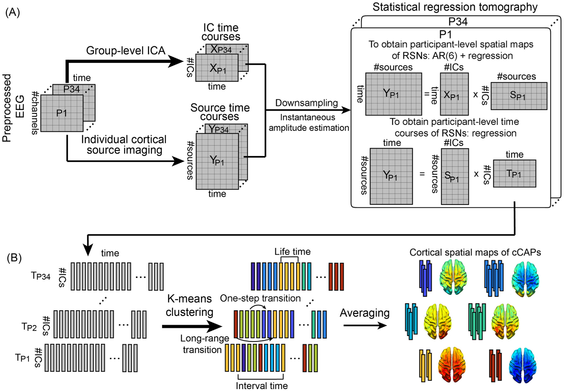
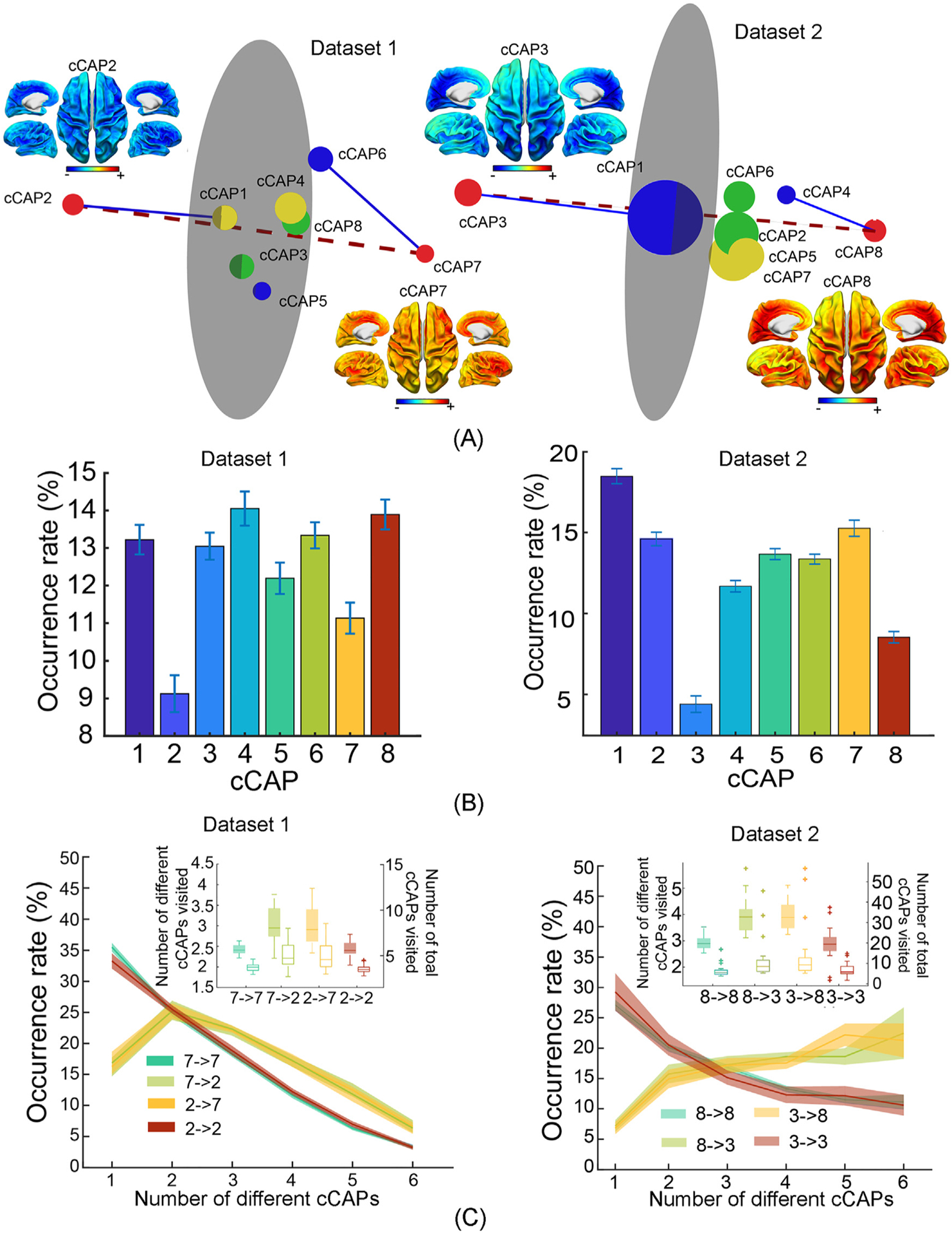
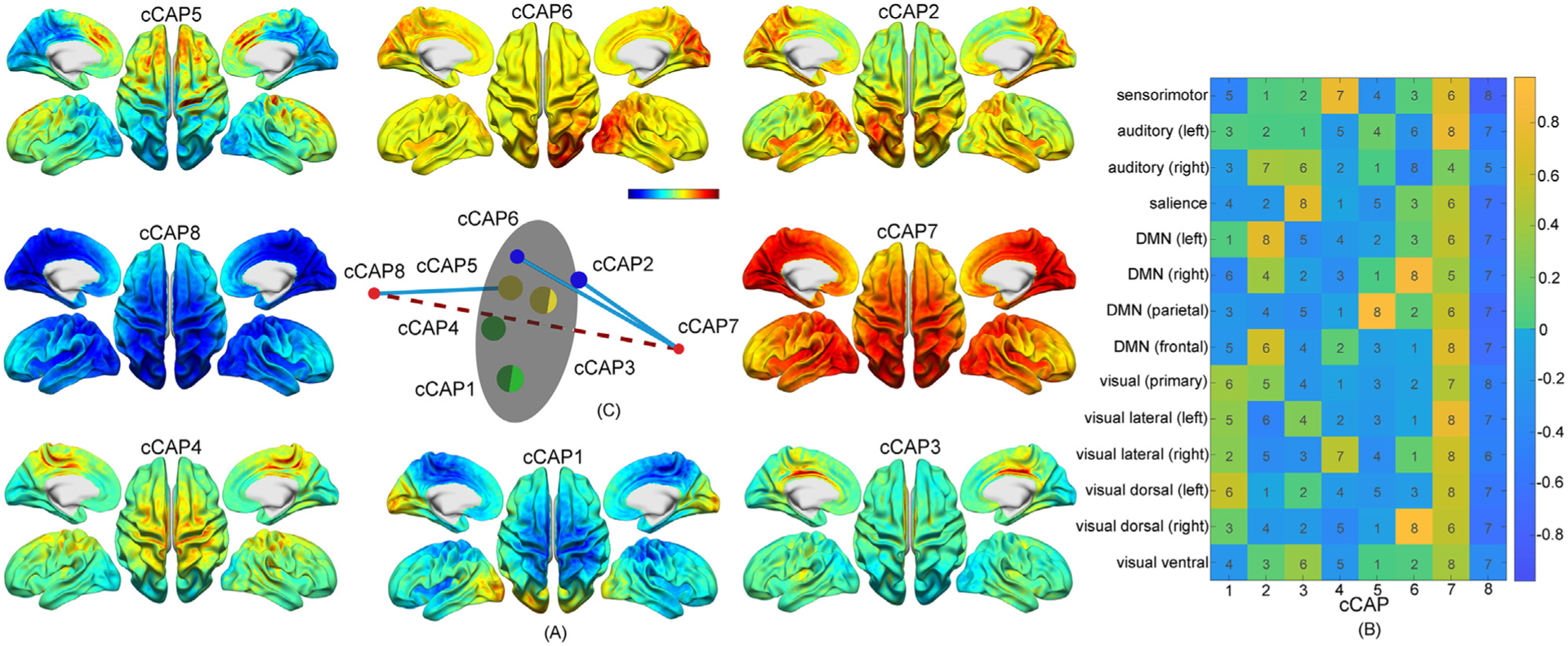
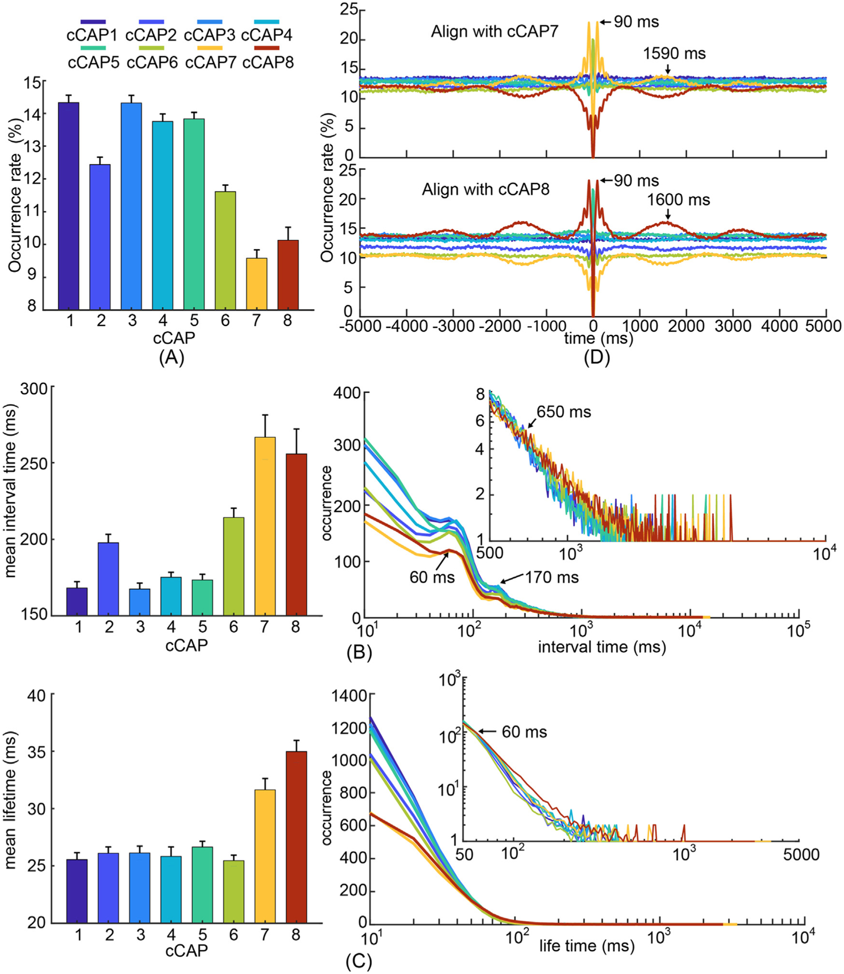
Spontaneous neural activity in human as assessed with resting-state functional magnetic resonance imaging (fMRI) exhibits brain-wide coordinated patterns in the frequency of < 0.1 Hz. However, understanding of fast brain-wide networks at the timescales of neuronal events (milliseconds to sub-seconds) and their spatial, spectral, and transitional characteristics remain limited due to the temporal constraints of hemodynamic signals. With milli-second resolution and whole-head coverage, scalp-based electroencephalography (EEG) provides a unique window into brain-wide networks with neuronal-timescale dynamics, shedding light on the organizing principles of brain functions. Using the state-of-the-art signal processing techniques, we reconstructed cortical neural tomography from resting-state EEG and extracted component-based co-activation patterns (cCAPs). These cCAPs revealed brain-wide intrinsic networks and their dynamics, indicating the configuration/reconfiguration of resting human brains into recurring and transitional functional states, which are featured with the prominent spatial phenomena of global patterns and anti-state pairs of co-(de)activations. Rich oscillational structures across a wide frequency band (i.e., 0.6 Hz, 5 Hz, and 10 Hz) were embedded in the nonstationary dynamics of these functional states. We further identified a superstructure that regulated between-state immediate and long-range transitions involving the entire set of identified cCAPs and governed a significant aspect of brain-wide network dynamics. These findings demonstrated how resting-state EEG data can be functionally decomposed using cCAPs to reveal rich dynamic structures of brain-wide human neural activations.
静息态功能磁共振成像 (fMRI) 评估的人类自发性神经活动在 < 0.1 Hz 的频率下表现出全脑协调模式。然而,由于血流动力学信号的时间限制,对神经元事件(毫秒到亚秒)时间尺度和其空间、谱和过渡特征的快速全脑网络的理解仍然有限。头皮脑电图 (EEG) 具有毫秒级分辨率和全头覆盖范围,为具有神经元时间尺度动力学的全脑网络提供了独特的窗口,揭示了大脑功能的组织原则。使用最先进的信号处理技术,我们从静息态 EEG 重建了皮质神经层析成像,并提取了基于组件的共激活模式 (cCAP)。这些 cCAP 揭示了全脑内在网络及其动力学,表明静息人脑会重新配置为反复出现和过渡的功能状态,其特点是全局模式和共(去)激活的反状态对的突出空间现象。在这些功能状态的非平稳动力学中嵌入了丰富的宽频带(即 0.6 Hz、5 Hz 和 10 Hz)的振荡结构。我们进一步确定了一个超级结构,该超级结构调节了涉及所有识别出的 cCAP 的状态间的即时和远程跃迁,并控制了全脑网络动力学的重要方面。这些发现表明,如何使用 cCAP 对静息态 EEG 数据进行功能分解,以揭示全脑人类神经激活的丰富动态结构。