• 文献检索
  • 文档翻译
  • 深度研究
  • 学术资讯
  • Suppr Zotero 插件Zotero 插件
  • 邀请有礼
  • 套餐&价格
  • 历史记录
应用&插件
Suppr Zotero 插件Zotero 插件浏览器插件Mac 客户端Windows 客户端微信小程序
定价
高级版会员购买积分包购买API积分包
服务
文献检索文档翻译深度研究API 文档MCP 服务
关于我们
关于 Suppr公司介绍联系我们用户协议隐私条款
关注我们

Suppr 超能文献

核心技术专利:CN118964589B侵权必究
粤ICP备2023148730 号-1Suppr @ 2026

文献检索

告别复杂PubMed语法,用中文像聊天一样搜索,搜遍4000万医学文献。AI智能推荐,让科研检索更轻松。

立即免费搜索

文件翻译

保留排版,准确专业,支持PDF/Word/PPT等文件格式,支持 12+语言互译。

免费翻译文档

深度研究

AI帮你快速写综述,25分钟生成高质量综述,智能提取关键信息,辅助科研写作。

立即免费体验

区分早期和晚期骨关节炎的T1 rho/T2映射弛豫时间的截断点。

Cutoff points of T1 rho/T2 mapping relaxation times distinguishing early-stage and advanced osteoarthritis.

作者信息

Yang Zhijian, Xie Chao, Ou Songwen, Zhao Minning, Lin Zhaowei

机构信息

Department of Joint Surgery, First Affiliated Hospital of Sun Yat-sen University, Guangzhou, Guangdong, China.

Department of Orthopaedics, Zhujiang Hospital, Southern Medical University, Guangzhou, Guangdong, China.

出版信息

Arch Med Sci. 2021 Aug 2;18(4):1004-1015. doi: 10.5114/aoms/140714. eCollection 2022.

DOI:10.5114/aoms/140714
PMID:35832709
原文链接:https://pmc.ncbi.nlm.nih.gov/articles/PMC9266714/
Abstract

INTRODUCTION

The histopathology grading system is the gold standard post-operative method to evaluate cartilage degeneration in knee osteoarthritis (OA). Magnetic resonance imaging (MRI) T1 rho/T2 mapping imaging can be used for preoperative detection. An association between histopathology and T1 rho/T2 mapping relaxation times was suggested in previous research. However, the cutoff point was not determined among different histopathology grades. Our study aimed to determine the cutoff point of T1 rho/T2 mapping.

MATERIAL AND METHODS

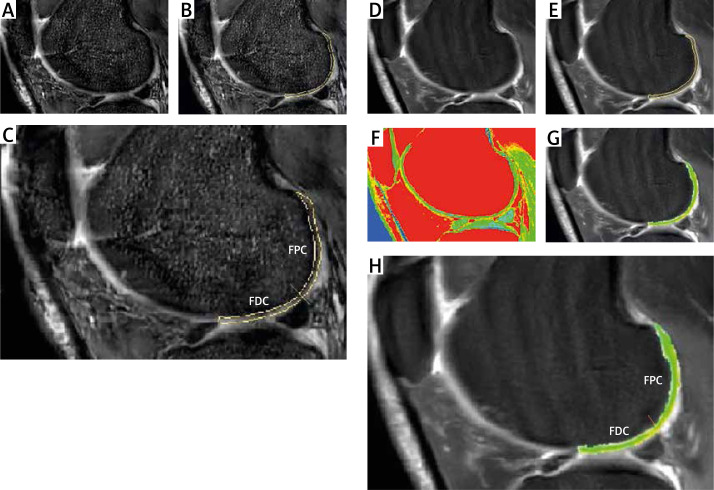
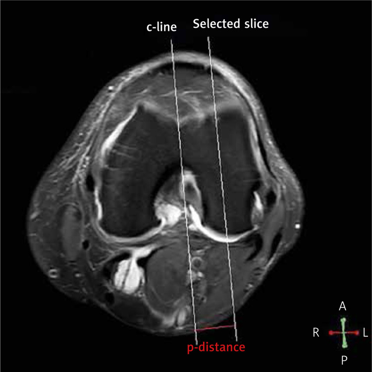
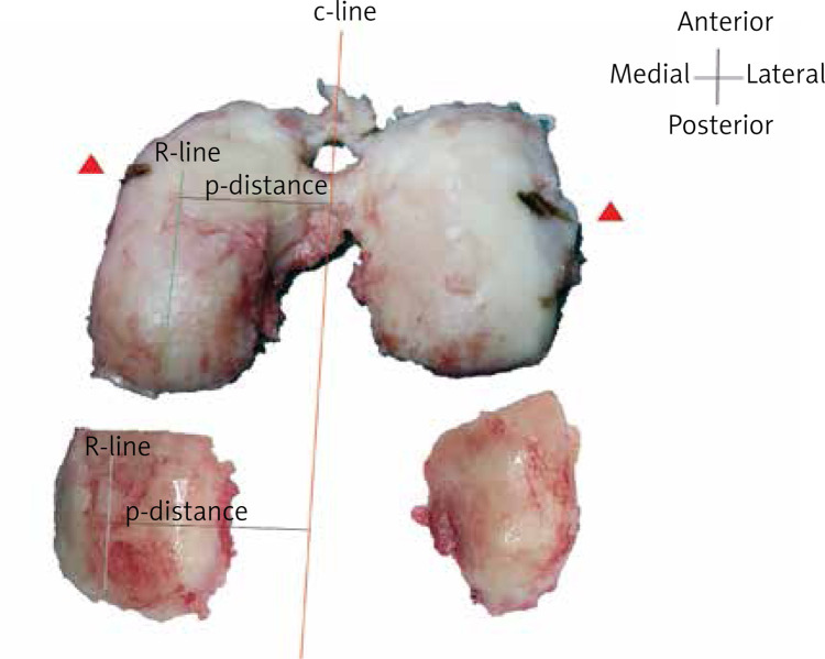
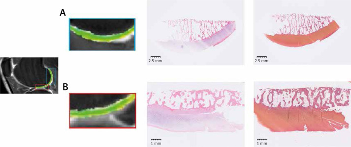
T1 rho/T2 mapping images were acquired from 80 samples before total knee replacements. Then the histopathology grading system was applied.

RESULTS

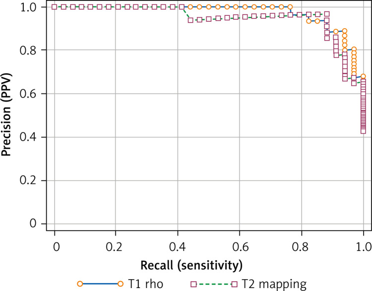
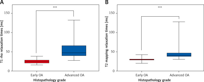
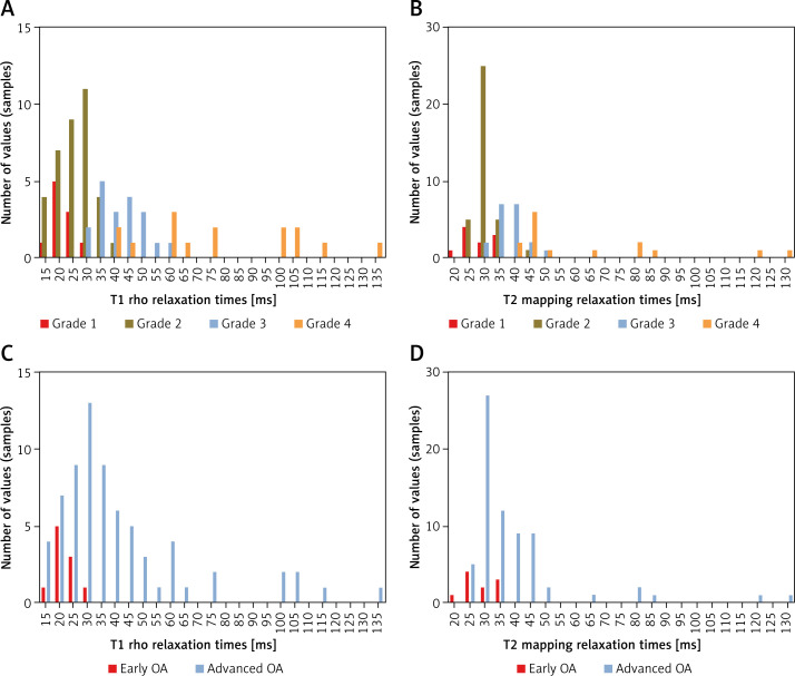
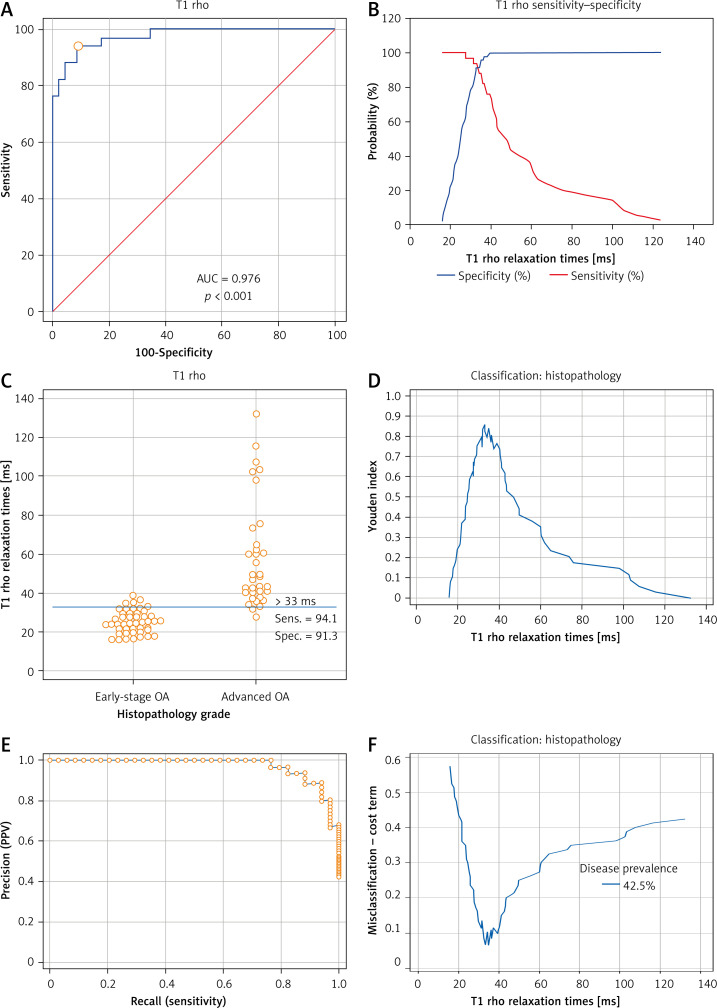
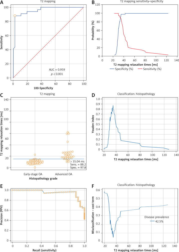
The mean T1 rho/T2 mapping relaxation times of 80 samples were 39.17 ms and 37.98 ms respectively. Significant differences were found in T1 rho/T2 mapping values between early-stage and advanced OA ( < 0.001). The cutoff point for T1 rho was 33 ms with a sensitivity of 94.12 (95% CI: 80-99.3) and a specificity of 91.30 (95% CI: 79.2-97.6). The cutoff point for T2 mapping was suggested as 35.04 ms with a sensitivity of 88.24 (95% CI: 72.5-96.7) and specificity of 97.83 (95% CI: 88.5-99.9). After bootstrap simulation, the 95% CI of the T1 rho/T2 mapping cutoff point was estimated as 29.36 to 36.32 ms and 34.8 to 35.04 ms respectively. The area under the PR curve of T1 rho/T2 mapping was 0.972 (95% CI: 0.925-0.992) and 0.949 (95% CI: 0.877-0.989) respectively.

CONCLUSIONS

The cutoff point of T1 rho relaxation times, which was suggested as 33 ms, could be used to distinguish early-stage and advanced OA.

摘要

引言

组织病理学分级系统是评估膝关节骨关节炎(OA)软骨退变的术后金标准方法。磁共振成像(MRI)T1ρ/T2映射成像可用于术前检测。先前的研究表明组织病理学与T1ρ/T2映射弛豫时间之间存在关联。然而,不同组织病理学分级之间的截断点尚未确定。我们的研究旨在确定T1ρ/T2映射的截断点。

材料与方法

在全膝关节置换术前从80个样本中获取T1ρ/T2映射图像。然后应用组织病理学分级系统。

结果

80个样本的平均T1ρ/T2映射弛豫时间分别为39.17毫秒和37.98毫秒。早期OA和晚期OA之间的T1ρ/T2映射值存在显著差异(<0.001)。T1ρ的截断点为33毫秒,灵敏度为94.12(95%CI:80-99.3),特异性为91.30(95%CI:79.2-97.6)。T2映射的截断点建议为35.04毫秒,灵敏度为88.24(95%CI:72.5-96.7),特异性为97.83(95%CI:88.5-99.9)。经过自助模拟,T1ρ/T2映射截断点的95%CI分别估计为29.36至36.32毫秒和34.8至35.04毫秒。T1ρ/T2映射的PR曲线下面积分别为0.972(95%CI:0.925-0.992)和0.949(95%CI:0.877-0.989)。

结论

T1ρ弛豫时间的截断点建议为33毫秒,可用于区分早期OA和晚期OA。

https://cdn.ncbi.nlm.nih.gov/pmc/blobs/7d9b/9266714/95be88dc277c/AMS-18-4-140714-g009.jpg
https://cdn.ncbi.nlm.nih.gov/pmc/blobs/7d9b/9266714/b99c5c56b3ac/AMS-18-4-140714-g001.jpg
https://cdn.ncbi.nlm.nih.gov/pmc/blobs/7d9b/9266714/0ad34d582c0a/AMS-18-4-140714-g002.jpg
https://cdn.ncbi.nlm.nih.gov/pmc/blobs/7d9b/9266714/13b7b8baba19/AMS-18-4-140714-g003.jpg
https://cdn.ncbi.nlm.nih.gov/pmc/blobs/7d9b/9266714/69ab247b1e16/AMS-18-4-140714-g004.jpg
https://cdn.ncbi.nlm.nih.gov/pmc/blobs/7d9b/9266714/5259457af588/AMS-18-4-140714-g005.jpg
https://cdn.ncbi.nlm.nih.gov/pmc/blobs/7d9b/9266714/341fca734b8b/AMS-18-4-140714-g006.jpg
https://cdn.ncbi.nlm.nih.gov/pmc/blobs/7d9b/9266714/1f15327bdf66/AMS-18-4-140714-g007.jpg
https://cdn.ncbi.nlm.nih.gov/pmc/blobs/7d9b/9266714/a334c01bbea6/AMS-18-4-140714-g008.jpg
https://cdn.ncbi.nlm.nih.gov/pmc/blobs/7d9b/9266714/95be88dc277c/AMS-18-4-140714-g009.jpg

相似文献

1
Cutoff points of T1 rho/T2 mapping relaxation times distinguishing early-stage and advanced osteoarthritis.区分早期和晚期骨关节炎的T1 rho/T2映射弛豫时间的截断点。
Arch Med Sci. 2021 Aug 2;18(4):1004-1015. doi: 10.5114/aoms/140714. eCollection 2022.
2
Associations of three-dimensional T1 rho MR mapping and three-dimensional T2 mapping with macroscopic and histologic grading as a biomarker for early articular degeneration of knee cartilage.三维 T1rho MR 映射与三维 T2 映射与宏观和组织学分级的相关性作为膝关节软骨早期关节退变的生物标志物。
Clin Rheumatol. 2017 Sep;36(9):2109-2119. doi: 10.1007/s10067-017-3645-2. Epub 2017 Apr 29.
3
The application of T1 and T2 relaxation time and magnetization transfer ratios to the early diagnosis of patellar cartilage osteoarthritis.T1 和 T2 弛豫时间和磁化传递率在髌股软骨骨关节炎早期诊断中的应用。
Skeletal Radiol. 2009 Nov;38(11):1055-62. doi: 10.1007/s00256-009-0769-8. Epub 2009 Aug 18.
4
The acute effect of running on knee articular cartilage and meniscus magnetic resonance relaxation times in young healthy adults.跑步对年轻健康成年人膝关节软骨和半月板磁共振弛豫时间的急性影响。
Am J Sports Med. 2012 Sep;40(9):2134-41. doi: 10.1177/0363546512449816. Epub 2012 Jun 22.
5
Meniscal measurements of T1rho and T2 at MR imaging in healthy subjects and patients with osteoarthritis.在健康受试者和骨关节炎患者中,通过磁共振成像测量半月板的T1rho和T2值。
Radiology. 2008 Nov;249(2):591-600. doi: 10.1148/radiol.2492071870.
6
Quantitative evaluation in combination with nonquantitative evaluation in early patellar cartilage osteoarthritis at 3.0 T.3.0T下早期髌股关节软骨骨关节炎的定量评估与非定量评估相结合
Clin Interv Aging. 2014 Jul 16;9:1133-43. doi: 10.2147/CIA.S65871. eCollection 2014.
7
In vivo biochemical 7.0 Tesla magnetic resonance: preliminary results of dGEMRIC, zonal T2, and T2* mapping of articular cartilage.体内生物化学7.0特斯拉磁共振成像:关节软骨延迟钆增强磁共振成像、分区T2及T2*成像的初步结果
Invest Radiol. 2008 Sep;43(9):619-26. doi: 10.1097/RLI.0b013e31817e9122.
8
An in vitro comparative study of T2 and T2* mappings of human articular cartilage at 3-Tesla MRI using histology as the standard of reference.一项以组织学为参考标准,在3特斯拉磁共振成像(MRI)下对人体关节软骨的T2和T2*映射进行的体外对比研究。
Skeletal Radiol. 2014 Jul;43(7):947-54. doi: 10.1007/s00256-014-1872-z. Epub 2014 Apr 9.
9
Quantitative imaging of anterior cruciate ligament (ACL) graft demonstrates longitudinal compositional changes and relationships with clinical outcomes at 2 years after ACL reconstruction.前交叉韧带(ACL)移植物的定量成像显示,在 ACL 重建 2 年后,其具有纵向组成变化,并与临床结果相关。
J Orthop Res. 2020 Jun;38(6):1289-1295. doi: 10.1002/jor.24572. Epub 2020 Jan 5.
10
T ρ and T mapping for the determination of articular cartilage denaturalization with osteonecrosis of the femoral head: A prospective controlled trial.基于 T ρ 和 T2 mapping 测定股骨头坏死伴有关节软骨退变的前瞻性对照研究
J Magn Reson Imaging. 2019 Mar;49(3):760-767. doi: 10.1002/jmri.26267. Epub 2018 Nov 21.

引用本文的文献

1
Evaluation of Cartilage Degeneration Using MRI T1ρ Mapping after Locomotion Training in Patients with Early Knee Osteoarthritis.早期膝关节骨关节炎患者运动训练后使用MRI T1ρ成像评估软骨退变情况
Prog Rehabil Med. 2025 Sep 5;10:20250020. doi: 10.2490/prm.20250020. eCollection 2025.
2
P13-516M14.1 Regulates Autophagy Through miR-429 via Both a ceRNA Network and Direct Interaction.P13 - 516M14.1通过ceRNA网络和直接相互作用经由miR - 429调控自噬。
Biochem Genet. 2025 May 28. doi: 10.1007/s10528-025-11143-x.
3
Synthetic Knee MRI T Maps as an Avenue for Clinical Translation of Quantitative Osteoarthritis Biomarkers.

本文引用的文献

1
Early Detection of Cartilage Degeneration: A Comparison of Histology, Fiber Bragg Grating-Based Micro-Indentation, and Atomic Force Microscopy-Based Nano-Indentation.早期软骨退变的检测:组织学、基于光纤布拉格光栅的微压痕和基于原子力显微镜的纳米压痕的比较。
Int J Mol Sci. 2020 Oct 6;21(19):7384. doi: 10.3390/ijms21197384.
2
L-Theanine Reduced the Development of Knee Osteoarthritis in Rats via Its Anti-Inflammation and Anti-Matrix Degradation Actions: In Vivo and In Vitro Study.L-茶氨酸通过其抗炎和抗基质降解作用减轻大鼠膝骨关节炎的发展:体内和体外研究。
Nutrients. 2020 Jul 3;12(7):1988. doi: 10.3390/nu12071988.
3
Quantitative measurement of T2, T1ρ and T1 relaxation times in articular cartilage and cartilage-bone interface by SE and UTE imaging at microscopic resolution.
合成膝关节MRI T图谱作为定量骨关节炎生物标志物临床转化的途径。
Bioengineering (Basel). 2023 Dec 24;11(1):17. doi: 10.3390/bioengineering11010017.
利用 SE 和 UTE 成像以微观分辨率定量测量关节软骨和软骨-骨界面的 T2、T1ρ 和 T1 弛豫时间。
J Magn Reson. 2018 Dec;297:76-85. doi: 10.1016/j.jmr.2018.10.008. Epub 2018 Oct 12.
4
Lutein prevents osteoarthritis through Nrf2 activation and downregulation of inflammation.叶黄素通过激活Nrf2和下调炎症反应来预防骨关节炎。
Arch Med Sci. 2018 Apr;14(3):617-624. doi: 10.5114/aoms.2016.59871. Epub 2016 May 12.
5
T1ρ/T2 mapping and histopathology of degenerative cartilage in advanced knee osteoarthritis.晚期膝关节骨关节炎中退变软骨的T1ρ/T2成像与组织病理学
World J Orthop. 2017 Apr 18;8(4):350-356. doi: 10.5312/wjo.v8.i4.350.
6
Associations of three-dimensional T1 rho MR mapping and three-dimensional T2 mapping with macroscopic and histologic grading as a biomarker for early articular degeneration of knee cartilage.三维 T1rho MR 映射与三维 T2 映射与宏观和组织学分级的相关性作为膝关节软骨早期关节退变的生物标志物。
Clin Rheumatol. 2017 Sep;36(9):2109-2119. doi: 10.1007/s10067-017-3645-2. Epub 2017 Apr 29.
7
Cluster analysis of quantitative MRI T and T relaxation times of cartilage identifies differences between healthy and ACL-injured individuals at 3T.对软骨的定量MRI T1和T2弛豫时间进行聚类分析,可识别出3T条件下健康个体与前交叉韧带损伤个体之间的差异。
Osteoarthritis Cartilage. 2017 Apr;25(4):513-520. doi: 10.1016/j.joca.2016.09.015. Epub 2016 Oct 5.
8
Functional MR Imaging Mapping of Human Articular Cartilage Response to Loading.功能性磁共振成像对人体关节软骨负重反应的映射。
Radiology. 2017 Feb;282(2):464-474. doi: 10.1148/radiol.2016160053. Epub 2016 Aug 26.
9
Early articular cartilage MRI T2 changes after anterior cruciate ligament reconstruction correlate with later changes in T2 and cartilage thickness.前交叉韧带重建术后早期关节软骨MRI T2改变与后期T2及软骨厚度改变相关。
J Orthop Res. 2017 Mar;35(3):699-706. doi: 10.1002/jor.23358. Epub 2016 Jul 29.
10
Early osteoarthritis of the knee.膝关节早期骨关节炎
Knee Surg Sports Traumatol Arthrosc. 2016 Jun;24(6):1753-62. doi: 10.1007/s00167-016-4068-3. Epub 2016 Mar 21.