Arellano-Orden E, Calero Acuña C, Sánchez-López V, López Ramírez C, Otero-Candelera R, Marín-Hinojosa C, López Campos Jl
Unidad Médico Quirúrgica de Enfermedades Respiratorias, Instituto de Biomedicina de Sevilla (IBIS), Hospital Universitario Virgen del Rocío/Universidad de SevillaUnidad Médico Quirúrgica de Enfermedades Respiratorias,Quirúrgica, Seville, Spain.
CIBER de Enfermedades Respiratorias (CIBERES), Instituto de Salud Carlos III, Madrid, Spain.
Eur Clin Respir J. 2022 Jul 8;9(1):2097377. doi: 10.1080/20018525.2022.2097377. eCollection 2022.
Airway epithelial cells and lung fibroblasts play an important role in the development of chronic lung disease, but the exact mechanisms responsible have not been clarified. Our objective was to investigate the involvement of these cells in the inflammatory response associated to chronic lung disease.
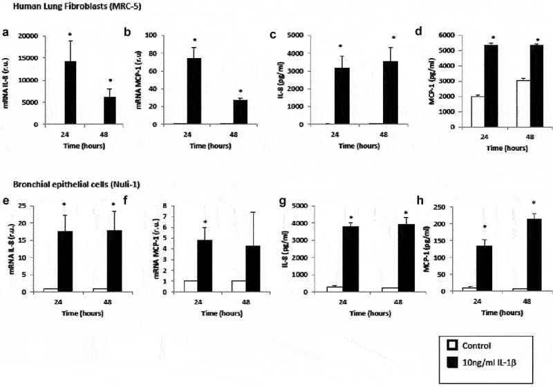
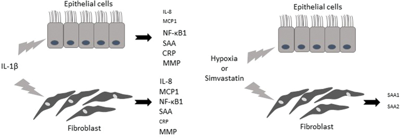
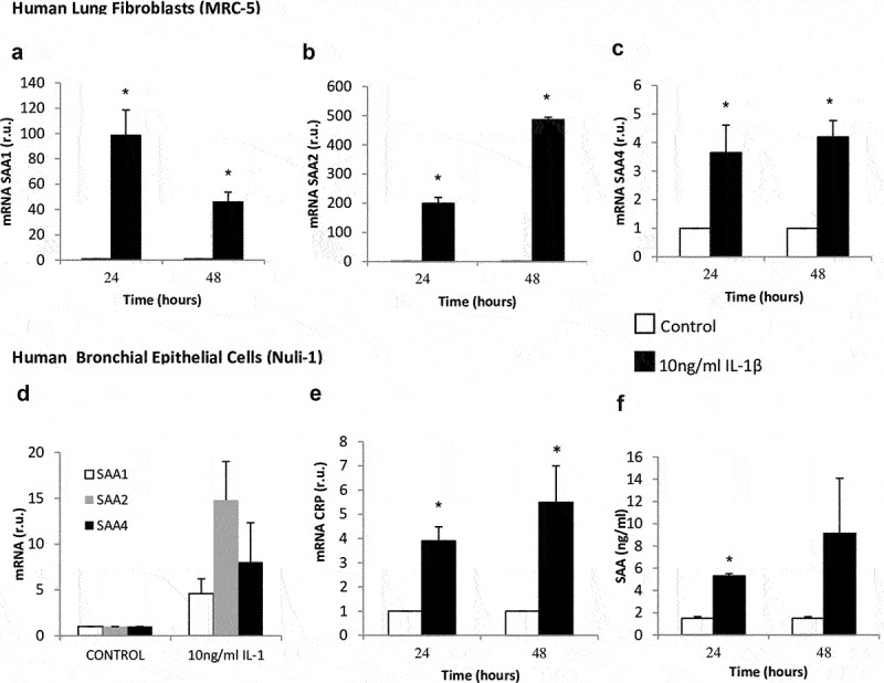
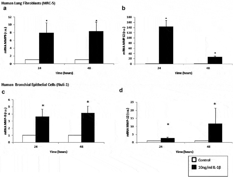
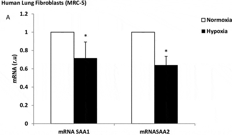
Human lung fibroblasts and airway epithelial cells were challenged with Interleukin-1β and hypoxia, and with inhibitory (simvastatin) stimuli of the inflammatory response. Expression of markers of local inflammation ((IL-8, monocyte chemoattractant protein-1 (MCP-1), factor-κB1 (NF-κB1)), systemic inflammation ((C-reactive protein (CRP) and serum amyloid A (SAA)) and proteases matrix metalloproteinase (MMP) 9 and 12 were assessed by PCR and ELISA. Apoptosis/necrosis was analyzed by flow cytometry.
Our results showed that the lung fibroblasts had a higher expression of local and systemic inflammation and protease activity markers when they were treated with IL-1β compared to airway epithelial cells. Under hypoxic conditions, we observed a decrease in systemic inflammation in lung fibroblasts, which was further attenuated by simvastatin.
The lung fibroblasts seem to be the main initially stimulated cells that could potentially trigger the inflammatory response, and be responsible for the eventual onset of chronic lung disease. The involvement of IL-1ß stimulation in systemic inflammatory and proteinase imbalance biomarkers is higher in lung fibroblasts. Apoptosis is not a predominant mechanism in these cells.
气道上皮细胞和肺成纤维细胞在慢性肺病的发展中起重要作用,但具体机制尚未阐明。我们的目的是研究这些细胞在与慢性肺病相关的炎症反应中的作用。
用人白细胞介素-1β和缺氧刺激人肺成纤维细胞和气道上皮细胞,并给予炎症反应的抑制性(辛伐他汀)刺激。通过聚合酶链反应(PCR)和酶联免疫吸附测定(ELISA)评估局部炎症标志物(白细胞介素-8、单核细胞趋化蛋白-1(MCP-1)、核因子-κB1(NF-κB1))、全身炎症标志物(C反应蛋白(CRP)和血清淀粉样蛋白A(SAA))以及基质金属蛋白酶(MMP)9和12的表达。通过流式细胞术分析细胞凋亡/坏死情况。
我们的结果表明,与气道上皮细胞相比,用白细胞介素-1β处理时,肺成纤维细胞中局部和全身炎症以及蛋白酶活性标志物的表达更高。在缺氧条件下,我们观察到肺成纤维细胞中全身炎症有所减轻,辛伐他汀可进一步减弱这种减轻。
肺成纤维细胞似乎是最初被刺激的主要细胞,可能引发炎症反应,并导致慢性肺病最终发病。白细胞介素-1β刺激在肺成纤维细胞的全身炎症和蛋白酶失衡生物标志物中的作用更大。细胞凋亡不是这些细胞中的主要机制。