Department of Ultrasound, Taizhou Hospital of Zhejiang Province Affiliated to Wenzhou Medical University, Taizhou 318053, Zhejiang, China.
Department of Thyroid and Breast Surgery, Taizhou Enze Medical Center (Group) Enze Hospital, Taizhou 318053, Zhejiang, China.
Contrast Media Mol Imaging. 2022 Jun 23;2022:3265342. doi: 10.1155/2022/3265342. eCollection 2022.
To investigate the significance of PAX8-PPAR expression in thyroid cancer and the application of a PAX8-PPAR-targeted ultrasound contrast agent in the early diagnosis of thyroid cancer.
In this study, the expression of PAX8-PPAR in thyroid cancer tissues, paracancer groups, and normal thyroid tissues was detected by western and immunohistochemical techniques; the effects of PAX8-PPAR expression inhibition on thyroid cancer cell growth, clonogenic ability, and antiapoptosis were examined. The terminal carboxylactic acid/hydroxyacetic acid copolymer (PLGA-COOH) nanoparticles were prepared by the double emulsification solvent volatilization method. The in vitro cytotoxicity of the targeted contrast agent was detected by MTS and other methods; LD50 was used to evaluate its short-term in vivo toxicity after intraperitoneal injection in mice.
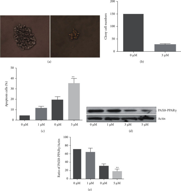
PAX8-PPAR expression was significantly increased in thyroid cancer tissues, and the expression level of PAX8-PPAR was closely correlated with TNM staging and lymph node metastasis ( < 0.05). In addition, PAX8-PPAR was also expressed at high levels in thyroid cancer cell lines relative to normal thyroid cells. MTS experiments showed that the PAX8-PPAR-targeted ultrasound nanocontrast agent had no significant toxic side effects on thyroid cells; countess observed that the contrast agent had no effect on cell survival and mortality; the LD50 assay showed that the targeted contrast agent had a wide safety range. Western blot showed the expression of caspase-3, BAX, and Bcl-2 in thyroid cancer cells, indicating that the nanocontrast agent has a good biosafety. In vitro targeting experiments showed that there were more nanospheres aggregated around the cells in the targeted contrast group. In vivo targeting imaging of nude mice revealed that the ultrasound signal was significantly enhanced in the targeted group compared with the nontargeted group after 20 min of LIFU irradiation.
PAX8-PPAR overexpression in thyroid cancer cell lines and thyroid cancer tissues promoted the proliferation and antiapoptotic ability of thyroid cancer cells and promoted the tumorigenic ability in nude mice in vivo. We successfully prepared a PAX8-PPAR-targeted ultrasound nanocontrast agent, which has regular morphology, uniform size, and high stability, and its liquid-gas phase change can be promoted at lower temperature. Therefore, this contrast agent can achieve US-targeted imaging and temperature phase transition function, and may have enhanced ultrasound imaging potential.
探讨 PAX8-PPAR 在甲状腺癌中的表达意义及 PAX8-PPAR 靶向超声造影剂在甲状腺癌早期诊断中的应用。
采用 Western blot 及免疫组化技术检测甲状腺癌组织、癌旁组织及正常甲状腺组织中 PAX8-PPAR 的表达,观察 PAX8-PPAR 表达抑制对甲状腺癌细胞生长、克隆形成及抗凋亡能力的影响。采用复乳溶剂挥发法制备端羧基聚乳酸-羟基乙酸共聚物(PLGA-COOH)纳米粒,通过 MTS 等方法检测靶向对比剂的体外细胞毒性;采用腹腔注射法检测其在小鼠体内的急性毒性(LD50)。
PAX8-PPAR 在甲状腺癌组织中呈高表达,且其表达水平与 TNM 分期及淋巴结转移密切相关( < 0.05)。此外,PAX8-PPAR 在甲状腺癌细胞系中也呈高表达,而在正常甲状腺细胞中低表达。MTS 实验显示,PAX8-PPAR 靶向超声纳米造影剂对甲状腺细胞无明显毒性作用;计数观察到该对比剂对细胞存活率和死亡率无影响;LD50 检测显示该靶向对比剂具有较宽的安全范围。Western blot 显示甲状腺癌细胞中 caspase-3、BAX、Bcl-2 表达增加,提示纳米造影剂具有良好的生物安全性。体外靶向实验显示,靶向组纳米球在细胞周围聚集较多。裸鼠体内靶向成像显示,LIFU 照射 20 min 后,靶向组的超声信号明显强于非靶向组。
PAX8-PPAR 在甲状腺癌细胞系和甲状腺癌组织中的高表达促进了甲状腺癌细胞的增殖和抗凋亡能力,并在体内促进了裸鼠的肿瘤生成能力。我们成功制备了 PAX8-PPAR 靶向超声纳米造影剂,其形态规则,粒径均匀,稳定性高,且其气-液相变可在较低温度下促进。因此,该对比剂可实现超声靶向成像及温度相变功能,可能具有增强超声成像的潜力。