Translational Medicine Collaborative Innovation Center, Shenzhen People's Hospital (The Second Clinical Medical College of Jinan University; The First Affiliated Hospital, Southern University of Science and Technology), Shenzhen, 518055, China.
Guangdong Engineering Technology Research Center of Stem Cell and Cell Therapy, Shenzhen Key Laboratory of Stem Cell Research and Clinical Transformation, Shenzhen, 518020, China.
Stem Cell Res Ther. 2022 Jul 15;13(1):314. doi: 10.1186/s13287-022-02996-9.
The biological activity and regenerative medicine of bone marrow mesenchymal stem cells (BMSCs) have been focal topics in the broad fields of diabetic wound repair. However, the molecular mechanisms are still largely elusive for other cellular processes that are regulated during BMSC treatment. Our previous studies have shown that hypoxia is not only a typical pathological phenomenon of wounds but also exerts a vital regulatory effect on cellular bioactivity. In this study, the beneficial effects of hypoxic BMSCs on the cellular behaviors of epidermal cells and diabetic wound healing were investigated.
The viability and secretion ability of hypoxic BMSCs were detected. The autophagy, proliferation and migration of HaCaT cells cultured with hypoxic BMSCs-derived conditioned medium were assessed by estimating the expression of autophagy-related proteins, MTS, EdU proliferation and scratch assays. And the role of the SMAD signaling pathway during hypoxic BMSC-evoked HaCaT cell autophagy was explored through a series of in vitro gain- and loss-of-function experiments. Finally, the therapeutic effects of hypoxic BMSCs were evaluated using full-thickness cutaneous diabetic wound model.
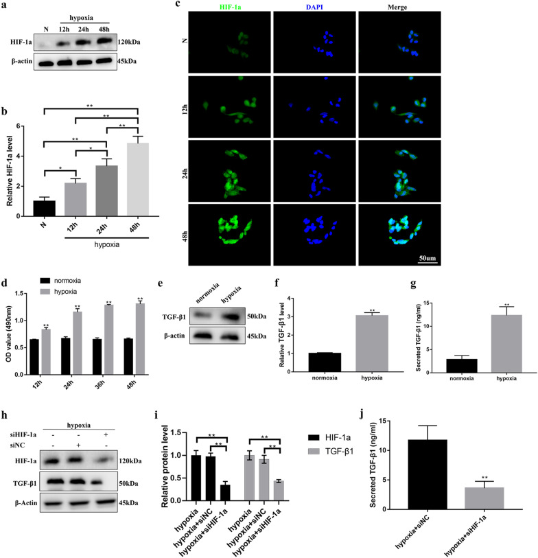
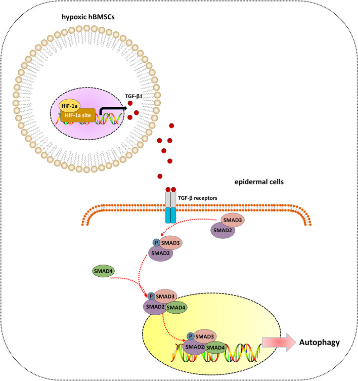
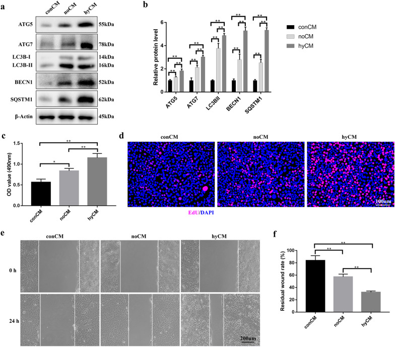
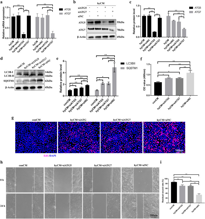
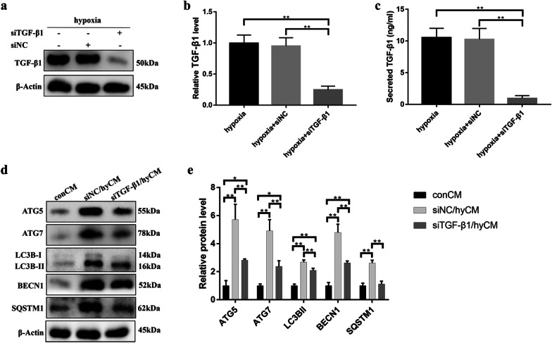
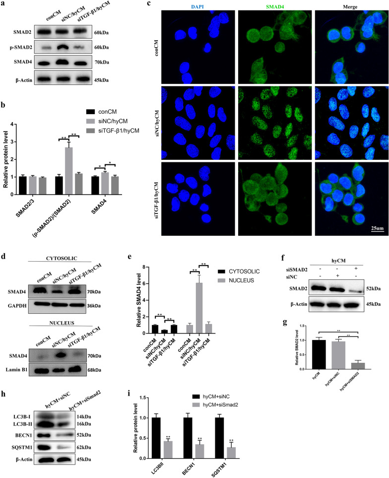
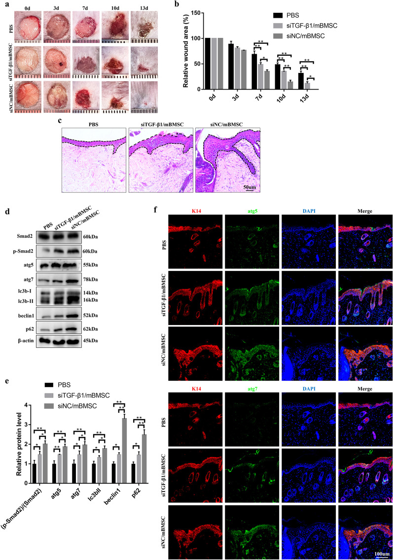
First, we demonstrated that hypoxic conditions intensify HIF-1α-mediated TGF-β1 secretion by BMSCs. Then, the further data revealed that BMSC-derived TGF-β1 was responsible for the activation of epidermal cell autophagy, which contributed to the induction of epidermal cell proliferation and migration. Here, the SMAD signaling pathway was identified as downstream of BMSC-derived TGF-β1 to regulate HaCaT cell autophagy. Moreover, the administration of BMSCs to diabetic wounds increased epidermal autophagy and the rate of re-epithelialization, leading to accelerated healing, and these effects were significantly attenuated, accompanied by the downregulation of Smad2 phosphorylation levels due to TGF-β1 interference in BMSCs.
In this report, we present evidence that uncovers a previously unidentified role of hypoxic BMSCs in regulating epidermal cell autophagy. The findings demonstrate that BMSC-based treatment by restoring epidermal cell autophagy could be an attractive therapeutic strategy for diabetic wounds and that the process is mediated by the HIF-1α/TGF-β1/SMAD pathway.
骨髓间充质干细胞(BMSCs)的生物学活性和再生医学一直是糖尿病创面修复等广泛领域的研究热点。然而,BMSC 治疗过程中其他受调控的细胞过程的分子机制仍很大程度上难以捉摸。我们之前的研究表明,缺氧不仅是伤口的一种典型病理现象,而且对细胞的生物活性也具有重要的调节作用。在这项研究中,我们研究了缺氧 BMSCs 对表皮细胞的细胞行为和糖尿病创面愈合的有益影响。
检测了缺氧 BMSCs 的活力和分泌能力。通过评估自噬相关蛋白的表达、MTS、EdU 增殖和划痕实验,评估了缺氧 BMSCs 条件培养基培养的 HaCaT 细胞的自噬、增殖和迁移。通过一系列体外功能获得和功能丧失实验,探讨了 SMAD 信号通路在缺氧 BMSC 诱导 HaCaT 细胞自噬过程中的作用。最后,利用全层皮肤糖尿病创面模型评估了缺氧 BMSCs 的治疗效果。
首先,我们证明了缺氧条件增强了 BMSCs 中 HIF-1α 介导的 TGF-β1 分泌。然后,进一步的数据表明,BMSC 衍生的 TGF-β1 负责激活表皮细胞自噬,从而促进表皮细胞的增殖和迁移。在这里,SMAD 信号通路被确定为 BMSC 衍生的 TGF-β1 下游调节 HaCaT 细胞自噬的途径。此外,BMSCs 给药到糖尿病创面增加了表皮自噬和再上皮化率,从而加速愈合,而由于 TGF-β1 干扰 BMSCs 中 Smad2 磷酸化水平,这些效果明显减弱。
在本报告中,我们提供了证据,揭示了缺氧 BMSCs 调节表皮细胞自噬的一个以前未被识别的作用。研究结果表明,通过恢复表皮细胞自噬的 BMSC 治疗可能是糖尿病创面的一种有吸引力的治疗策略,该过程是由 HIF-1α/TGF-β1/SMAD 途径介导的。