• 文献检索
  • 文档翻译
  • 深度研究
  • 学术资讯
  • Suppr Zotero 插件Zotero 插件
  • 邀请有礼
  • 套餐&价格
  • 历史记录
应用&插件
Suppr Zotero 插件Zotero 插件浏览器插件Mac 客户端Windows 客户端微信小程序
定价
高级版会员购买积分包购买API积分包
服务
文献检索文档翻译深度研究API 文档MCP 服务
关于我们
关于 Suppr公司介绍联系我们用户协议隐私条款
关注我们

Suppr 超能文献

核心技术专利:CN118964589B侵权必究
粤ICP备2023148730 号-1Suppr @ 2026

文献检索

告别复杂PubMed语法,用中文像聊天一样搜索,搜遍4000万医学文献。AI智能推荐,让科研检索更轻松。

立即免费搜索

文件翻译

保留排版,准确专业,支持PDF/Word/PPT等文件格式,支持 12+语言互译。

免费翻译文档

深度研究

AI帮你快速写综述,25分钟生成高质量综述,智能提取关键信息,辅助科研写作。

立即免费体验

新生儿脑室内出血后的继发性脑损伤:纤毛室管膜的作用。

Secondary Brain Injury Following Neonatal Intraventricular Hemorrhage: The Role of the Ciliated Ependyma.

作者信息

Dawes William

机构信息

Alder Hey Children's Hospital, Liverpool, United Kingdom.

NIHR Great Ormond Street Hospital BRC, London, United Kingdom.

出版信息

Front Pediatr. 2022 Jun 30;10:887606. doi: 10.3389/fped.2022.887606. eCollection 2022.

DOI:10.3389/fped.2022.887606
PMID:35844746
原文链接:https://pmc.ncbi.nlm.nih.gov/articles/PMC9280684/
Abstract

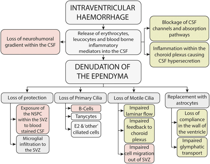
Intraventricular hemorrhage is recognized as a leading cause of hydrocephalus in the developed world and a key determinant of neurodevelopmental outcome following premature birth. Even in the absence of haemorrhagic infarction or posthaemorrhagic hydrocephalus, there is increasing evidence of neuropsychiatric and neurodevelopmental sequelae. The pathophysiology underlying this injury is thought to be due to a primary destructive and secondary developmental insult, but the exact mechanisms remain elusive and this has resulted in a paucity of therapeutic interventions. The presence of blood within the cerebrospinal fluid results in the loss of the delicate neurohumoral gradient within the developing brain, adversely impacting on the tightly regulated temporal and spatial control of cell proliferation and migration of the neural stem progenitor cells within the subventricular zone. In addition, haemolysis of the erythrocytes, associated with the release of clotting factors and leucocytes into the cerebrospinal (CSF), results in a toxic and inflammatory CSF microenvironment which is harmful to the periventricular tissues, resulting in damage and denudation of the multiciliated ependymal cells which line the choroid plexus and ventricular system. The ependyma plays a critical role in the developing brain and beyond, acting as both a protector and gatekeeper to the underlying parenchyma, controlling influx and efflux across the CSF to brain interstitial fluid interface. In this review I explore the hypothesis that damage and denudation of the ependymal layer at this critical juncture in the developing brain, seen following IVH, may adversely impact on the brain microenvironment, exposing the underlying periventricular tissues to toxic and inflammatory CSF, further exacerbating disordered activity within the subventricular zone (SVZ). By understanding the impact that intraventricular hemorrhage has on the microenvironment within the CSF, and the consequences that this has on the multiciliated ependymal cells which line the neuraxis, we can begin to develop and test novel therapeutic interventions to mitigate damage and reduce the associated morbidity.

摘要

在发达国家,脑室内出血被认为是脑积水的主要原因,也是早产后脑神经发育结局的关键决定因素。即使没有出血性梗死或出血后脑积水,越来越多的证据表明存在神经精神和神经发育后遗症。这种损伤的病理生理学被认为是由于原发性破坏和继发性发育损伤,但确切机制仍然难以捉摸,这导致治疗干预措施匮乏。脑脊液中存在血液会导致发育中的大脑内微妙的神经体液梯度丧失,对脑室下区神经干祖细胞增殖和迁移的严格时空控制产生不利影响。此外,红细胞溶血,伴随着凝血因子和白细胞释放到脑脊液中,会导致有毒和炎症性的脑脊液微环境,对脑室周围组织有害,导致衬于脉络丛和脑室系统的多纤毛室管膜细胞受损和剥脱。室管膜在发育中的大脑及其他方面起着关键作用,既是潜在实质的保护者和守门人,控制脑脊液与脑间质液界面的流入和流出。在这篇综述中,我探讨了这样一种假说,即脑室内出血后在发育中的大脑这个关键节点上室管膜层的损伤和剥脱,可能会对脑微环境产生不利影响,使潜在的脑室周围组织暴露于有毒和炎症性脑脊液中,进一步加剧脑室下区(SVZ)内的紊乱活动。通过了解脑室内出血对脑脊液微环境的影响,以及这对沿神经轴排列的多纤毛室管膜细胞的影响,我们可以开始开发和测试新的治疗干预措施,以减轻损伤并降低相关发病率。

https://cdn.ncbi.nlm.nih.gov/pmc/blobs/1e71/9280684/f847578dd59d/fped-10-887606-g0004.jpg
https://cdn.ncbi.nlm.nih.gov/pmc/blobs/1e71/9280684/7312610cd5d1/fped-10-887606-g0001.jpg
https://cdn.ncbi.nlm.nih.gov/pmc/blobs/1e71/9280684/580549eb68f2/fped-10-887606-g0002.jpg
https://cdn.ncbi.nlm.nih.gov/pmc/blobs/1e71/9280684/0fb6336c118e/fped-10-887606-g0003.jpg
https://cdn.ncbi.nlm.nih.gov/pmc/blobs/1e71/9280684/f847578dd59d/fped-10-887606-g0004.jpg

相似文献

1
Secondary Brain Injury Following Neonatal Intraventricular Hemorrhage: The Role of the Ciliated Ependyma.新生儿脑室内出血后的继发性脑损伤:纤毛室管膜的作用。
Front Pediatr. 2022 Jun 30;10:887606. doi: 10.3389/fped.2022.887606. eCollection 2022.
2
Neuroblast proliferation on the surface of the adult rat striatal wall after focal ependymal loss by intracerebroventricular injection of neuraminidase.通过脑室内注射神经氨酸酶导致局灶性室管膜缺失后,成年大鼠纹状体壁表面的神经母细胞增殖。
J Comp Neurol. 2008 Apr 1;507(4):1571-87. doi: 10.1002/cne.21618.
3
High-Speed Video Microscopy of Ependymal Cilia in Brain Organotypic and Cell Culture Models.高速视频显微镜在脑器官型和细胞培养模型中的室管膜纤毛研究
Methods Mol Biol. 2024;2725:239-250. doi: 10.1007/978-1-0716-3507-0_15.
4
Ventricular Zone Disruption in Human Neonates With Intraventricular Hemorrhage.患有脑室内出血的人类新生儿的脑室区破坏
J Neuropathol Exp Neurol. 2017 May 1;76(5):358-375. doi: 10.1093/jnen/nlx017.
5
Inflammation of the choroid plexus and ependymal layer of the ventricle following intraventricular hemorrhage.脑室出血后脉络丛和脑室管膜层的炎症。
Transl Stroke Res. 2011 Jun;2(2):227-31. doi: 10.1007/s12975-011-0070-8.
6
Extracellular hemoglobin - mediator of inflammation and cell death in the choroid plexus following preterm intraventricular hemorrhage.细胞外血红蛋白——早产儿脑室内出血后脉络丛炎症和细胞死亡的介质
J Neuroinflammation. 2014 Dec 2;11:200. doi: 10.1186/s12974-014-0200-9.
7
Neurospheres from neural stem/neural progenitor cells (NSPCs) of non-hydrocephalic HTx rats produce neurons, astrocytes and multiciliated ependyma: the cerebrospinal fluid of normal and hydrocephalic rats supports such a differentiation.来自非脑积水 HTx 大鼠的神经干/神经祖细胞 (NSPCs) 的神经球产生神经元、星形胶质细胞和多纤毛室管膜:正常和脑积水大鼠的脑脊液支持这种分化。
Cell Tissue Res. 2018 Aug;373(2):421-438. doi: 10.1007/s00441-018-2828-8. Epub 2018 Apr 12.
8
Human Cord Blood Derived Unrestricted Somatic Stem Cells Restore Aquaporin Channel Expression, Reduce Inflammation and Inhibit the Development of Hydrocephalus After Experimentally Induced Perinatal Intraventricular Hemorrhage.人脐带血来源的无限制体细胞干细胞可恢复水通道蛋白通道表达,减轻炎症并抑制实验性诱导的围产期脑室内出血后脑积水的发展。
Front Cell Neurosci. 2021 Apr 9;15:633185. doi: 10.3389/fncel.2021.633185. eCollection 2021.
9
NLRP3 inflammasome-mediated choroid plexus hypersecretion contributes to hydrocephalus after intraventricular hemorrhage via phosphorylated NKCC1 channels.NLRP3 炎性小体介导体脉络丛过度分泌导致脑室出血后继发脑积水通过磷酸化 NKCC1 通道。
J Neuroinflammation. 2022 Jun 21;19(1):163. doi: 10.1186/s12974-022-02530-x.
10
Glioblastoma disrupts the ependymal wall and extracellular matrix structures of the subventricular zone.胶质母细胞瘤破坏脑室下区的室管膜壁和细胞外基质结构。
Fluids Barriers CNS. 2022 Jul 11;19(1):58. doi: 10.1186/s12987-022-00354-8.

引用本文的文献

1
Transcriptomic and histological characteristics of innate immune activation in brain parenchyma in a rat model of neonatal intraventricular hemorrhage.新生大鼠脑室内出血模型脑实质内固有免疫激活的转录组学和组织学特征
Fluids Barriers CNS. 2025 Jul 3;22(1):68. doi: 10.1186/s12987-025-00678-1.
2
Research priorities for non-invasive therapies to improve hydrocephalus outcomes.改善脑积水治疗效果的非侵入性疗法的研究重点。
Fluids Barriers CNS. 2025 Mar 3;22(1):24. doi: 10.1186/s12987-025-00632-1.
3
Incidence of Communicating Hydrocephalus Following Intraventricular Hemorrhage Among Adult Patients Treated at a Hospital in Jeddah, Saudi Arabia: A Retrospective Study.

本文引用的文献

1
New insights into the management of post-hemorrhagic hydrocephalus.对出血后脑积水管理的新认识。
Semin Perinatol. 2022 Aug;46(5):151597. doi: 10.1016/j.semperi.2022.151597. Epub 2022 Mar 12.
2
Periventricular- intraventricular hemorrhage in the premature infant- A historical perspective.早产儿脑室周围-脑室内出血-历史视角。
Semin Perinatol. 2022 Aug;46(5):151591. doi: 10.1016/j.semperi.2022.151591. Epub 2022 Mar 12.
3
Neurodevelopmental outcomes of premature infants with intraventricular hemorrhage across a lifespan.
沙特阿拉伯吉达一家医院治疗的成年患者脑室内出血后交通性脑积水的发生率:一项回顾性研究
Cureus. 2025 Jan 20;17(1):e77699. doi: 10.7759/cureus.77699. eCollection 2025 Jan.
4
Extrauterine support of pre-term lambs achieves similar transcriptomic profiling to late pre-term lamb brains.早产羔羊的宫外支持可实现与晚期早产羔羊脑相似的转录组特征。
Sci Rep. 2024 Nov 21;14(1):28840. doi: 10.1038/s41598-024-79095-7.
5
Iron homeostasis and post-hemorrhagic hydrocephalus: a review.铁稳态与出血后脑积水:综述
Front Neurol. 2024 Jan 12;14:1287559. doi: 10.3389/fneur.2023.1287559. eCollection 2023.
6
High-Speed Video Microscopy of Ependymal Cilia in Brain Organotypic and Cell Culture Models.高速视频显微镜在脑器官型和细胞培养模型中的室管膜纤毛研究
Methods Mol Biol. 2024;2725:239-250. doi: 10.1007/978-1-0716-3507-0_15.
7
Therapeutic strategies to recover ependymal barrier after inflammatory damage: relevance for recovering neurogenesis during development.炎症损伤后恢复室管膜屏障的治疗策略:对发育过程中神经发生恢复的相关性。
Front Neurosci. 2023 Jun 15;17:1204197. doi: 10.3389/fnins.2023.1204197. eCollection 2023.
8
The Role of Oxidative Stress in the Progression of Secondary Brain Injury Following Germinal Matrix Hemorrhage.氧化应激在脑室内出血后继发性脑损伤进展中的作用。
Transl Stroke Res. 2024 Jun;15(3):647-658. doi: 10.1007/s12975-023-01147-3. Epub 2023 Mar 17.
早产儿脑室出血的全生命周期神经发育结局。
Semin Perinatol. 2022 Aug;46(5):151594. doi: 10.1016/j.semperi.2022.151594. Epub 2022 Mar 12.
4
Biochemical profile of human infant cerebrospinal fluid in intraventricular hemorrhage and post-hemorrhagic hydrocephalus of prematurity.人类婴儿脑室出血和早产儿出血后脑积水的脑脊液生化特征。
Fluids Barriers CNS. 2021 Dec 24;18(1):62. doi: 10.1186/s12987-021-00295-8.
5
A clinical primer for the glymphatic system.淋巴系统临床入门指南。
Brain. 2022 Apr 29;145(3):843-857. doi: 10.1093/brain/awab428.
6
Post-haemorrhagic hydrocephalus is associated with poorer surgical and neurodevelopmental sequelae than other causes of infant hydrocephalus.出血后脑积水比其他婴儿脑积水的病因与更差的手术和神经发育后遗症相关。
Childs Nerv Syst. 2021 Nov;37(11):3385-3396. doi: 10.1007/s00381-021-05226-4. Epub 2021 Jun 19.
7
Ependymal ciliary motion and their role in congenital hydrocephalus.室管膜纤毛的运动及其在先天性脑积水形成中的作用。
Childs Nerv Syst. 2021 Nov;37(11):3355-3364. doi: 10.1007/s00381-021-05194-9. Epub 2021 May 17.
8
Intraventricular haemorrhage and posthaemorrhagic ventricular dilatation: moving beyond CSF diversion.脑室内出血和出血后脑室扩张:超越脑脊液引流。
Childs Nerv Syst. 2021 Nov;37(11):3375-3383. doi: 10.1007/s00381-021-05206-8. Epub 2021 May 15.
9
White matter injury in infants with intraventricular haemorrhage: mechanisms and therapies.脑室出血婴儿的脑白质损伤:发病机制与治疗策略。
Nat Rev Neurol. 2021 Apr;17(4):199-214. doi: 10.1038/s41582-020-00447-8. Epub 2021 Jan 27.
10
Timing of Temporizing Neurosurgical Treatment in Relation to Shunting and Neurodevelopmental Outcomes in Posthemorrhagic Ventricular Dilatation of Prematurity: A Meta-analysis.早产儿出血后脑积水分流与神经发育结局相关的暂时机能性神经外科治疗时机:一项荟萃分析。
J Pediatr. 2021 Jul;234:54-64.e20. doi: 10.1016/j.jpeds.2021.01.030. Epub 2021 Jan 21.