Ning Lin, Geng Yang, Lovett-Barron Matthew, Niu Xiaoman, Deng Mengying, Wang Liang, Ataie Niloufar, Sens Alex, Ng Ho-Leung, Chen Shoudeng, Deisseroth Karl, Lin Michael Z, Chu Jun
Department of Neurobiology, Stanford University, Stanford, CA, United States.
Interdisciplinary Research Center on Biology and Chemistry, Shanghai Institute of Organic Chemistry, Chinese Academy of Sciences, China University of Chinese Academy of Sciences, Beijing, China.
Front Cell Dev Biol. 2022 Jun 29;10:893468. doi: 10.3389/fcell.2022.893468. eCollection 2022.
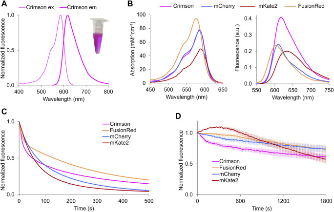
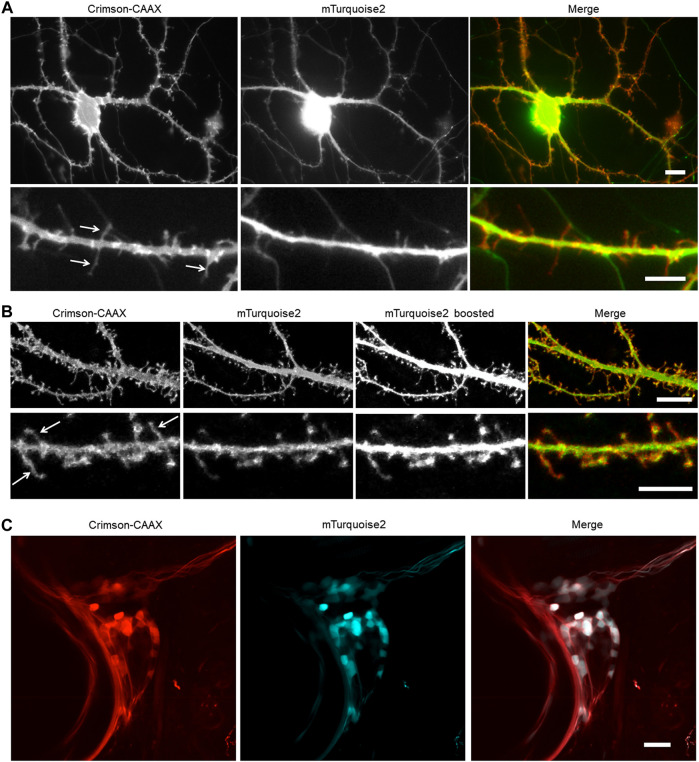
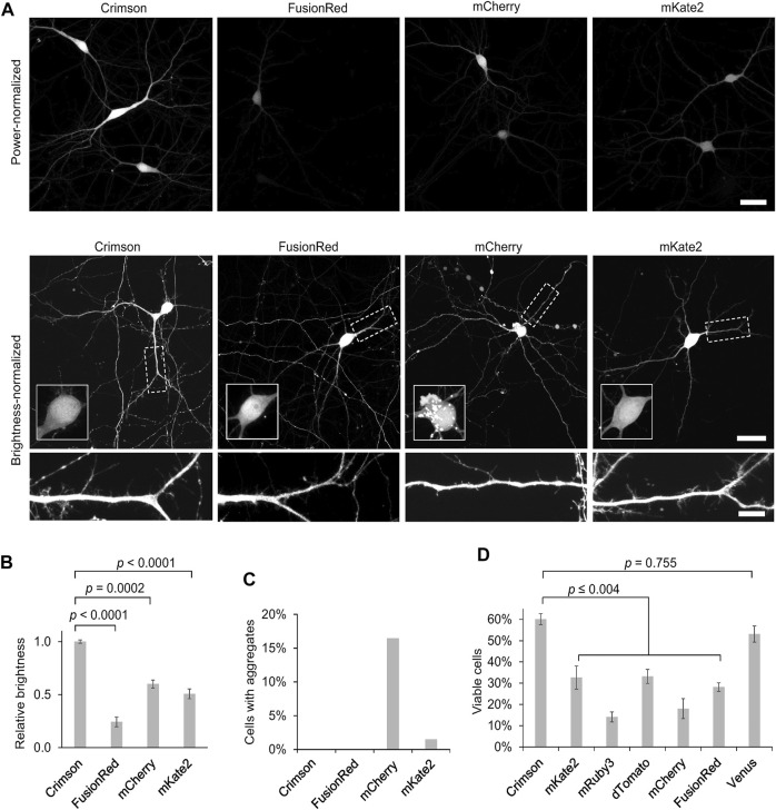
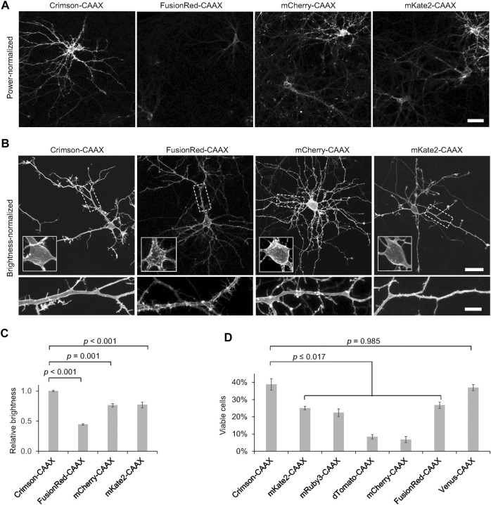
Red fluorescent proteins are useful as morphological markers in neurons, often complementing green fluorescent protein-based probes of neuronal activity. However, commonly used red fluorescent proteins show aggregation and toxicity in neurons or are dim. We report the engineering of a bright red fluorescent protein, Crimson, that enables long-term morphological labeling of neurons without aggregation or toxicity. Crimson is similar to mCherry and mKate2 in fluorescence spectra but is 100 and 28% greater in molecular brightness, respectively. We used a membrane-localized Crimson-CAAX to label thin neurites, dendritic spines and filopodia, enhancing detection of these small structures compared to cytosolic markers.
红色荧光蛋白作为神经元中的形态标记物很有用,常作为基于绿色荧光蛋白的神经元活动探针的补充。然而,常用的红色荧光蛋白在神经元中会出现聚集和毒性,或者亮度较低。我们报告了一种亮红色荧光蛋白Crimson的工程改造,它能够对神经元进行长期形态标记,而不会出现聚集或毒性。Crimson在荧光光谱上与mCherry和mKate2相似,但分子亮度分别高出100%和28%。我们使用膜定位的Crimson-CAAX来标记细神经突、树突棘和丝状伪足,与胞质标记物相比,增强了对这些小结构的检测。