Ophthalmology Unit, Mental Health, Neurosciences, and Sense Organs (NESMOS) Department, Faculty of Medicine and Psychology, University of Rome Sapienza, Rome, Italy.
St. Andrea Hospital, Via di Grottarossa 1035/1039, 00189, Rome, Italy.
Graefes Arch Clin Exp Ophthalmol. 2023 Feb;261(2):291-301. doi: 10.1007/s00417-022-05743-1. Epub 2022 Jul 19.
To provide a review of the literature on oculodermal melanocytosis (ODM) with a focus on the diagnostic and therapeutic implications of multimodal imaging techniques in the management of ophthalmic complications.
The authors carried out a literature search on PubMed, Medline, and Scopus of English language articles published on ODM through August 2021. This review presents traditional and novel diagnostic methods in the diagnosis and follow-up of patients with particular emphasis on addressing the role of imaging in the management of the ophthalmic complications of the condition towards improving current practice patterns.
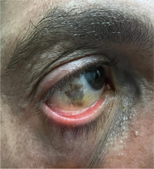
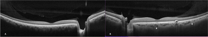
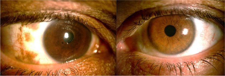
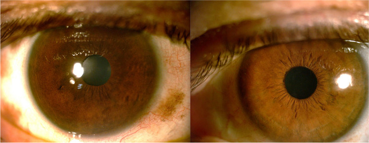
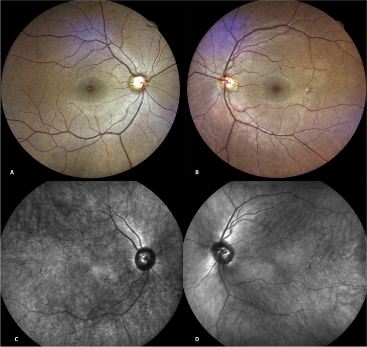
ODM is a rare, prevalently unilateral, congenital condition that presents with brown or blue/gray flat asymptomatic lesions of the skin, mucosae, episclera/sclera, and uvea localized within the territory of distribution of the ophthalmic and mandibular branches of the trigeminal nerve. Glaucoma and predisposition to uveal melanoma are the main ophthalmic complications. Diagnosis and management are through comprehensive opthalmological examination and traditional imaging methods such as ultrasonography and fluorescein/indocyanine green angiography as pigmentation of the fundus can conceal subtle retinal and choroidal alterations. Anterior segment optical coherence tomography and ultrasound biomicroscopy are used to evaluate the anterior segment and the ciliary body in the presence of glaucoma or melanoma of the anterior uveal tract. Fundus autofluorescence and retinal pigment epithelium (RPE) alterations are of aid in the differential diagnosis between choroidal nevi and melanoma. Enhanced depth imaging spectral domain optical coherence tomography offers outstanding in vivo evaluation of the dimensions and details of tumors or nevi and surrounding choroidal tissues and small choroidal melanomas may show distortions of the retinal and sub-retinal profile, presence of intra and sub-retinal fluid, abnormalities of the RPE, and compression of the choriocapillaris.
Novel multimodal imaging techniques are significant in the diagnosis and management of the ophthalmic complications of ODM. Fundus autofluorescence and enhanced depth spectral domain optical coherence tomography have adjunctive value in the detection of early-stage melanoma and differential diagnosis between nevi and melanoma. Awareness of current and emerging imaging techniques can propagate improved standardized definition and assessment of the complications of ODM.
对眼皮肤黑素细胞增多症(ODM)的文献进行综述,重点介绍多模态成像技术在眼部并发症管理中的诊断和治疗意义。
作者对 2021 年 8 月前在 PubMed、Medline 和 Scopus 上发表的关于 ODM 的英文文献进行了检索。本综述介绍了传统和新型诊断方法,特别强调了成像在处理该疾病眼部并发症中的作用,以改善当前的实践模式。
ODM 是一种罕见的、普遍单侧的先天性疾病,表现为皮肤、黏膜、巩膜/球结膜和葡萄膜的棕色或蓝/灰色扁平无症状病变,位于三叉神经眼支和下颌支的支配区域内。青光眼和葡萄膜黑色素瘤易感性是主要的眼部并发症。诊断和治疗通过全面的眼科检查和传统的成像方法,如超声检查和荧光素/吲哚青绿血管造影,因为眼底的色素沉着可能掩盖细微的视网膜和脉络膜改变。前节光学相干断层扫描和超声生物显微镜用于评估眼前节和睫状体,以评估青光眼或眼前节葡萄膜黑色素瘤的存在。眼底自发荧光和视网膜色素上皮(RPE)改变有助于鉴别脉络膜痣和黑色素瘤。增强深度成像光谱域光学相干断层扫描可对肿瘤或痣及其周围脉络膜组织的尺寸和细节进行出色的体内评估,小的脉络膜黑色素瘤可能表现为视网膜和视网膜下轮廓的扭曲、视网膜内和视网膜下积液、RPE 异常和脉络膜毛细血管压缩。
新型多模态成像技术对 ODM 的眼部并发症的诊断和管理具有重要意义。眼底自发荧光和增强深度光谱域光学相干断层扫描在检测早期黑色素瘤和鉴别痣和黑色素瘤方面具有附加价值。了解当前和新兴的成像技术可以促进对 ODM 并发症的标准化定义和评估。