The Wellcome Centre for Cell-Matrix Research and.
Blond McIndoe Laboratories, University of Manchester, Manchester Academic Health Science Centre, Manchester, United Kingdom.
JCI Insight. 2022 Aug 22;7(16):e156115. doi: 10.1172/jci.insight.156115.
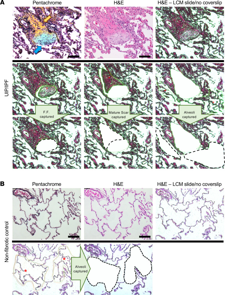
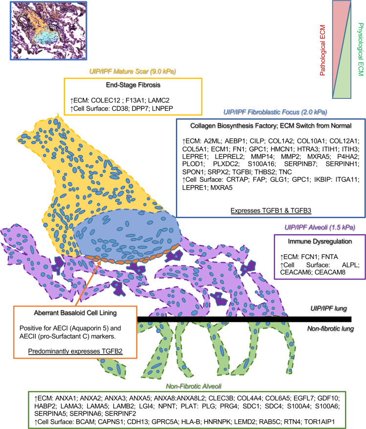
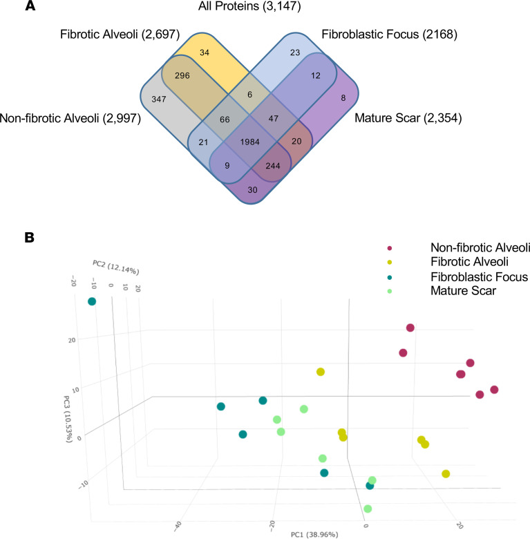
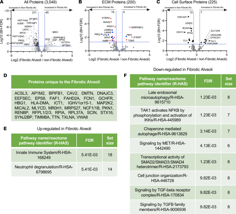
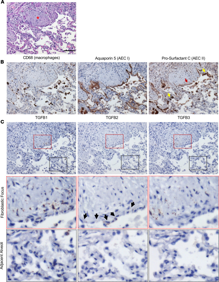
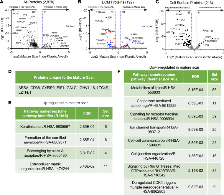
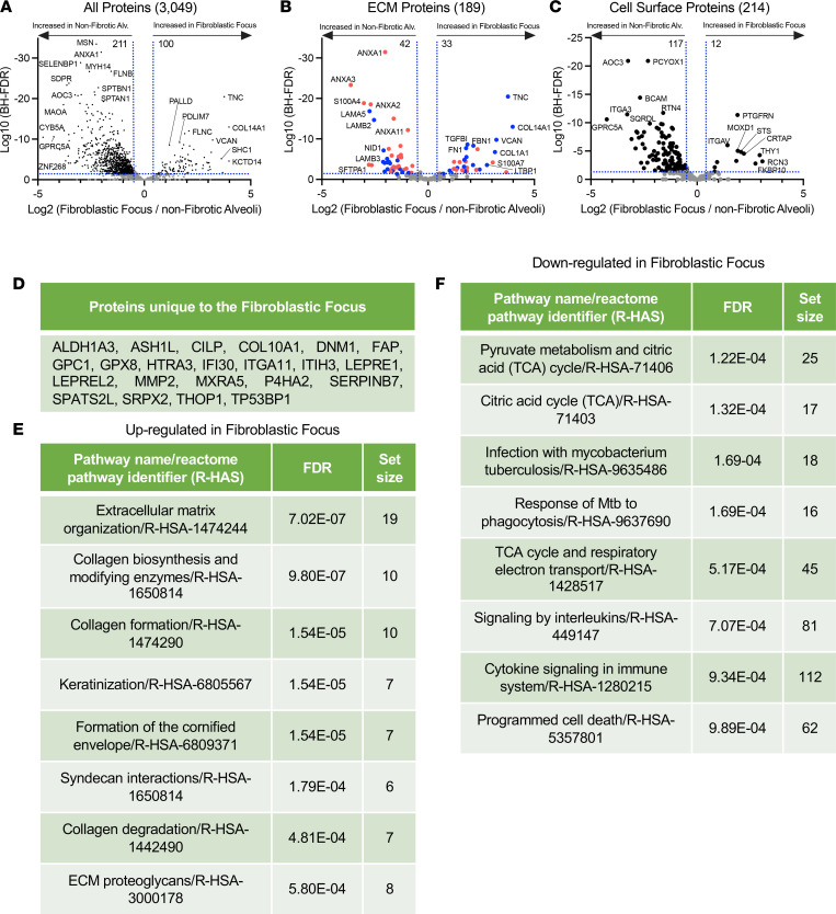
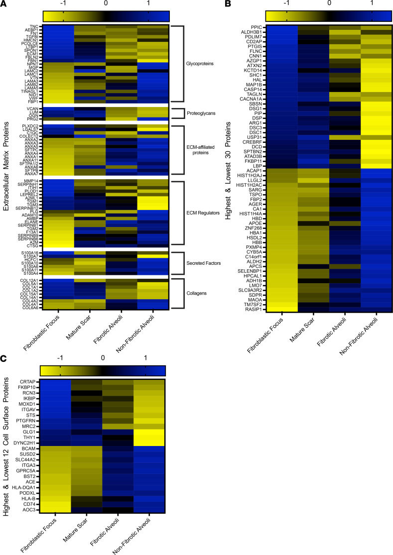
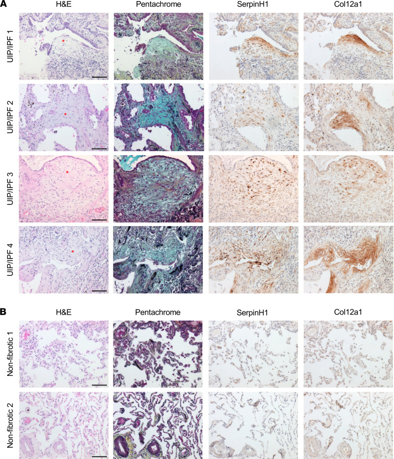
Usual interstitial pneumonia (UIP) is a histological pattern characteristic of idiopathic pulmonary fibrosis (IPF). The UIP pattern is patchy with histologically normal lung adjacent to dense fibrotic tissue. At this interface, fibroblastic foci (FF) are present and are sites where myofibroblasts and extracellular matrix (ECM) accumulate. Utilizing laser capture microdissection-coupled mass spectrometry, we interrogated the FF, adjacent mature scar, and adjacent alveoli in 6 fibrotic (UIP/IPF) specimens plus 6 nonfibrotic alveolar specimens as controls. The data were subjected to qualitative and quantitative analysis and histologically validated. We found that the fibrotic alveoli protein signature is defined by immune deregulation as the strongest category. The fibrotic mature scar classified as end-stage fibrosis whereas the FF contained an overabundance of a distinctive ECM compared with the nonfibrotic control. Furthermore, FF were positive for both TGFB1 and TGFB3, whereas the aberrant basaloid cell lining of FF was predominantly positive for TGFB2. In conclusion, spatial proteomics demonstrated distinct protein compositions in the histologically defined regions of UIP/IPF tissue. These data revealed that FF are the main site of collagen biosynthesis and that the adjacent alveoli are abnormal. This essential information will inform future mechanistic studies on fibrosis progression.
普通型间质性肺炎(UIP)是特发性肺纤维化(IPF)的一种组织学模式。UIP 模式呈斑片状,相邻的正常肺组织与致密的纤维组织相邻。在这个界面上,存在成纤维细胞灶(FF),是肌成纤维细胞和细胞外基质(ECM)积聚的部位。利用激光捕获显微切割-质谱联用技术,我们对 6 例纤维化(UIP/IPF)标本中的 FF、相邻成熟瘢痕和相邻肺泡以及 6 例非纤维化肺泡标本进行了检测。对数据进行了定性和定量分析,并进行了组织学验证。我们发现,纤维化肺泡的蛋白特征是免疫失调,这是最强的类别。纤维化成熟瘢痕被归类为终末期纤维化,而 FF 中含有大量独特的 ECM,与非纤维化对照相比明显过多。此外,FF 均为 TGFB1 和 TGFB3 阳性,而 FF 的基底样细胞衬里主要为 TGFB2 阳性。总之,空间蛋白质组学显示 UIP/IPF 组织中不同的组织学定义区域存在不同的蛋白质组成。这些数据表明,FF 是胶原生物合成的主要部位,而相邻的肺泡是异常的。这些重要信息将为纤维化进展的未来机制研究提供信息。