Wang Yunyang, Wang Mo, Liu Yunshan, Tao Hui, Banerjee Somesh, Srinivasan Shanthi, Nemeth Elizabeta, Czaja Mark J, He Peijian
Division of Digestive Diseases, Department of Medicine, Emory University School of Medicine, Atlanta, GA, USA.
Division of Digestive Diseases, Department of Medicine, Emory University School of Medicine, Atlanta, GA, USA; Gastroenterology Research, Atlanta VA Health Care System, Decatur, GA, USA.
Redox Biol. 2022 Sep;55:102407. doi: 10.1016/j.redox.2022.102407. Epub 2022 Jul 14.
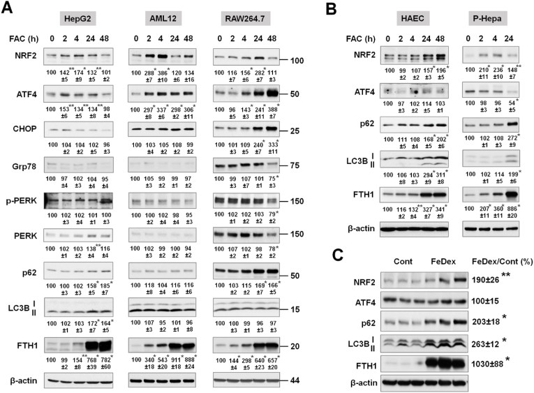
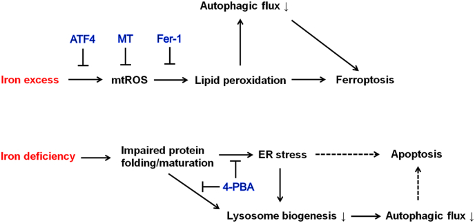
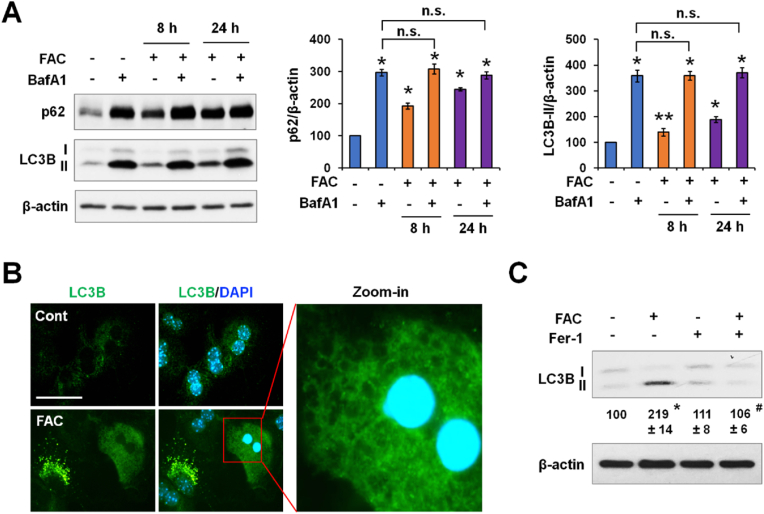
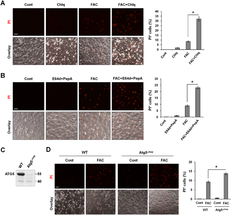
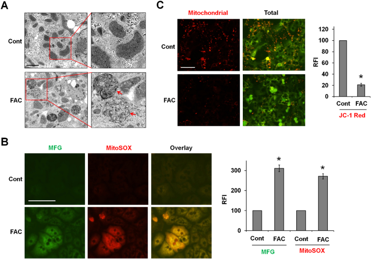
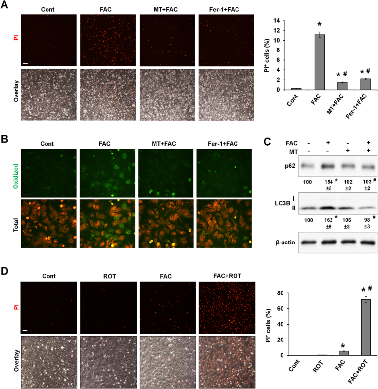
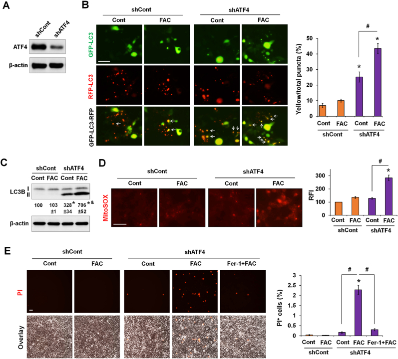
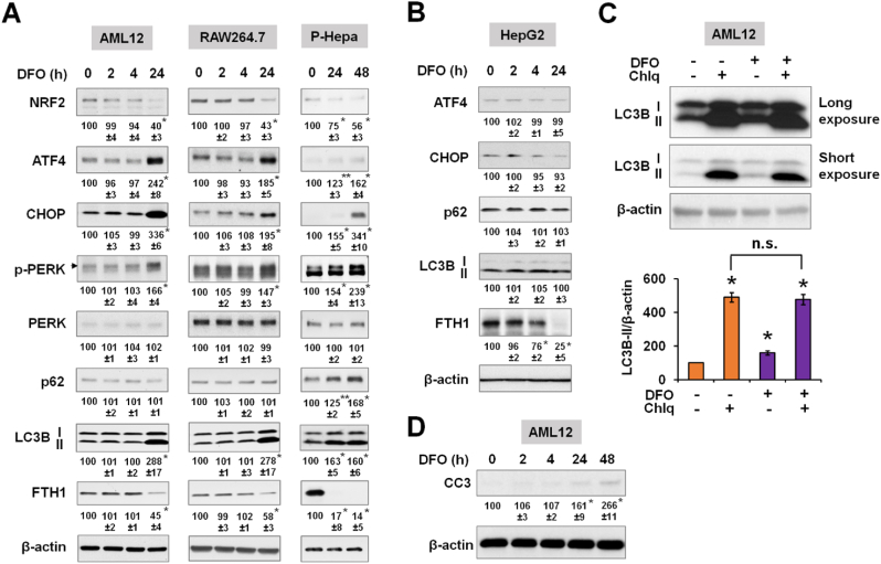
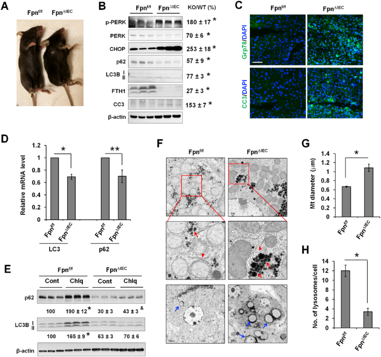
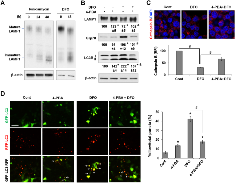
Iron is a mineral essential for blood production and a variety of critical cellular functions. Altered iron metabolism has been increasingly observed in many diseases and disorders, but a comprehensive and mechanistic understanding of the cellular impact of impaired iron metabolism is still lacking. We examined the effects of iron overload or iron deficiency on cellular stress responses and autophagy which collectively regulate cell homeostasis and survival. Acute iron loading led to increased mitochondrial ROS (mtROS) production and damage, lipid peroxidation, impaired autophagic flux, and ferroptosis. Iron-induced mtROS overproduction is the mechanism of increased lipid peroxidation, impaired autophagy, and the induction of ferroptosis. Iron excess-induced ferroptosis was cell-type dependent and regulated by activating transcription factor 4 (ATF4). Upregulation of ATF4 mitigated iron-induced autophagic dysfunction and ferroptosis, whereas silencing of ATF4 expression impaired autophagy and resulted in increased mtROS production and ferroptosis. Employing autophagy-deficient hepatocytes and different autophagy inhibitors, we further showed that autophagic impairment sensitized cells to iron-induced ferroptosis. In contrast, iron deficiency activated the endoplasmic reticulum (ER) stress response, decreased autophagy, and induced apoptosis. Decreased autophagy associated with iron deficiency was due to ER stress, as reduction of ER stress by 4-phenylbutyric acid (4-PBA) improved autophagic flux. The mechanism of decreased autophagy in iron deficiency is a disruption in lysosomal biogenesis due to impaired posttranslational maturation of lysosomal membrane proteins. In conclusion, iron excess and iron deficiency cause different forms of cell stress and death in part through the common mechanism of impaired autophagic function.
铁是一种对血液生成和多种关键细胞功能必不可少的矿物质。在许多疾病和病症中,铁代谢改变的情况日益常见,但对铁代谢受损的细胞影响仍缺乏全面而深入的机制性理解。我们研究了铁过载或铁缺乏对细胞应激反应和自噬的影响,这些共同调节着细胞内环境稳定和细胞存活。急性铁负荷导致线粒体活性氧(mtROS)生成增加和损伤、脂质过氧化、自噬流受损以及铁死亡。铁诱导的mtROS过度生成是脂质过氧化增加、自噬受损和铁死亡诱导的机制。铁过量诱导的铁死亡具有细胞类型依赖性,并由激活转录因子4(ATF4)调节。ATF4的上调减轻了铁诱导的自噬功能障碍和铁死亡,而ATF4表达的沉默则损害了自噬并导致mtROS生成增加和铁死亡。利用自噬缺陷型肝细胞和不同的自噬抑制剂,我们进一步表明自噬损伤使细胞对铁诱导的铁死亡敏感。相比之下,铁缺乏激活了内质网(ER)应激反应,降低了自噬,并诱导了细胞凋亡。与铁缺乏相关的自噬减少是由于内质网应激,因为4-苯基丁酸(4-PBA)减轻内质网应激改善了自噬流。铁缺乏时自噬减少的机制是由于溶酶体膜蛋白翻译后成熟受损导致溶酶体生物发生受到破坏。总之,铁过量和铁缺乏部分通过自噬功能受损这一共同机制导致不同形式的细胞应激和死亡。