• 文献检索
  • 文档翻译
  • 深度研究
  • 学术资讯
  • Suppr Zotero 插件Zotero 插件
  • 邀请有礼
  • 套餐&价格
  • 历史记录
应用&插件
Suppr Zotero 插件Zotero 插件浏览器插件Mac 客户端Windows 客户端微信小程序
定价
高级版会员购买积分包购买API积分包
服务
文献检索文档翻译深度研究API 文档MCP 服务
关于我们
关于 Suppr公司介绍联系我们用户协议隐私条款
关注我们

Suppr 超能文献

核心技术专利:CN118964589B侵权必究
粤ICP备2023148730 号-1Suppr @ 2026

文献检索

告别复杂PubMed语法,用中文像聊天一样搜索,搜遍4000万医学文献。AI智能推荐,让科研检索更轻松。

立即免费搜索

文件翻译

保留排版,准确专业,支持PDF/Word/PPT等文件格式,支持 12+语言互译。

免费翻译文档

深度研究

AI帮你快速写综述,25分钟生成高质量综述,智能提取关键信息,辅助科研写作。

立即免费体验

三维动脉瘤壁增强与症状表现相关。

3D aneurysm wall enhancement is associated with symptomatic presentation.

机构信息

Department of Neurology, The University of Iowa Hospitals and Clinics, Iowa City, Iowa, USA.

Institute for Clinical and Translational Science, The University of Iowa, Iowa City, Iowa, USA.

出版信息

J Neurointerv Surg. 2023 Aug;15(8):747-752. doi: 10.1136/jnis-2022-019125. Epub 2022 Jul 19.

DOI:10.1136/jnis-2022-019125
PMID:35853699
原文链接:https://pmc.ncbi.nlm.nih.gov/articles/PMC10173164/
Abstract

BACKGROUND

Aneurysm wall enhancement (AWE) is a potential surrogate biomarker for aneurysm instability. Previous studies have assessed AWE using 2D multiplanar methods, most of which were conducted qualitatively.

OBJECTIVE

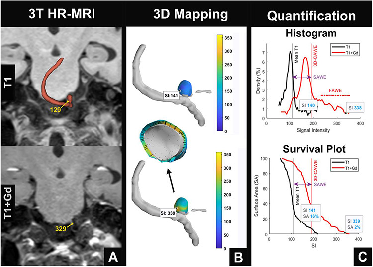
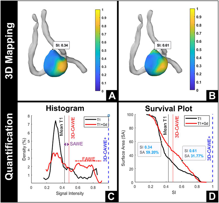
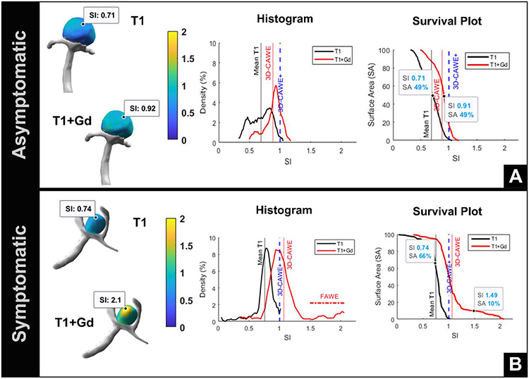
To use a new quantitative tool to analyze a large cohort of saccular aneurysms with 3D-AWE maps METHODS: Saccular aneurysms were imaged prospectively with 3T high resolution MRI. 3D-AWE maps of symptomatic (defined as ruptured or presentation with sentinel headache/cranial nerve neuropathy) and asymptomatic aneurysms were created by extending orthogonal probes from the aneurysm lumen into the wall. Three metrics were used to characterize enhancement: 3D circumferential AWE (3D-CAWE), aneurysm-specific contrast uptake (SAWE), and focal AWE (FAWE). Aneurysms with a circumferential AWE higher than the corpus callosum (3D-CAWE ≥1) were classified as 3D-CAWE+. Symptomatic presentation was analyzed with univariate and multivariate logistic models. Aneurysm size, size ratio, aspect ratio, irregular morphology, and PHASES and ELAPSS scores were compared with the new AWE metrics. Bleb and microhemorrhage analyses were also performed.

RESULTS

Ninety-three aneurysms were analyzed. 3D-CAWE, SAWE, and FAWE were associated with symptomatic status (OR=1.34, 1.25, and 1.08, respectively). A multivariate model including aneurysm size, 3D-CAWE+, age, female gender, and FAWE detected symptomatic status with 80% specificity and 90% sensitivity (area under the curve=0.914, =0.967). FAWE was also associated with irregular morphology and high-risk location (p=0.043 and p=0.001, respectively). In general, blebs enhanced 56% more than the aneurysm body. Areas of microhemorrhage co-localized with areas of increased SAWE (p=0.047).

CONCLUSIONS

3D-AWE mapping provides a new set of metrics that could potentially improve the identification of symptomatic aneurysms.

摘要

背景

动脉瘤壁增强(AWE)是动脉瘤不稳定的潜在替代生物标志物。先前的研究使用二维多平面方法评估了 AWE,其中大多数方法都是定性的。

目的

使用新的定量工具分析大量囊状动脉瘤的 3D-AWE 图谱。

方法

使用 3T 高分辨率 MRI 前瞻性地对囊状动脉瘤进行成像。通过从动脉瘤管腔向壁延伸正交探针,创建有症状(定义为破裂或出现先兆性头痛/颅神经神经病)和无症状动脉瘤的 3D-AWE 图谱。使用三个指标来描述增强:三维周向 AWE(3D-CAWE)、动脉瘤特异性对比摄取(SAWE)和局灶性 AWE(FAWE)。将周向 AWE 高于胼胝体(3D-CAWE≥1)的动脉瘤归类为 3D-CAWE+。使用单变量和多变量逻辑模型分析症状表现。比较了动脉瘤大小、大小比、纵横比、不规则形态以及 PHASES 和 ELAPSS 评分与新的 AWE 指标。还进行了微泡和微出血分析。

结果

分析了 93 个动脉瘤。3D-CAWE、SAWE 和 FAWE 与症状状态相关(OR=1.34、1.25 和 1.08)。包括动脉瘤大小、3D-CAWE+、年龄、女性性别和 FAWE 的多变量模型检测到症状状态的特异性为 80%,敏感性为 90%(曲线下面积=0.914,=0.967)。FAWE 还与不规则形态和高风险位置相关(p=0.043 和 p=0.001)。一般来说,微泡比动脉瘤体增强 56%。微出血区域与 SAWE 增加区域共定位(p=0.047)。

结论

3D-AWE 图谱提供了一组新的指标,可能有助于提高对有症状动脉瘤的识别。

https://cdn.ncbi.nlm.nih.gov/pmc/blobs/8b2f/10173164/40bd0637411a/nihms-1893955-f0003.jpg
https://cdn.ncbi.nlm.nih.gov/pmc/blobs/8b2f/10173164/46d11ea15d78/nihms-1893955-f0001.jpg
https://cdn.ncbi.nlm.nih.gov/pmc/blobs/8b2f/10173164/3b637a4c5d76/nihms-1893955-f0002.jpg
https://cdn.ncbi.nlm.nih.gov/pmc/blobs/8b2f/10173164/40bd0637411a/nihms-1893955-f0003.jpg
https://cdn.ncbi.nlm.nih.gov/pmc/blobs/8b2f/10173164/46d11ea15d78/nihms-1893955-f0001.jpg
https://cdn.ncbi.nlm.nih.gov/pmc/blobs/8b2f/10173164/3b637a4c5d76/nihms-1893955-f0002.jpg
https://cdn.ncbi.nlm.nih.gov/pmc/blobs/8b2f/10173164/40bd0637411a/nihms-1893955-f0003.jpg

相似文献

1
3D aneurysm wall enhancement is associated with symptomatic presentation.三维动脉瘤壁增强与症状表现相关。
J Neurointerv Surg. 2023 Aug;15(8):747-752. doi: 10.1136/jnis-2022-019125. Epub 2022 Jul 19.
2
Association of circumferential aneurysm wall enhancement with recurrence after coiling of unruptured intracranial aneurysms: a preliminary vessel wall imaging study.未破裂颅内动脉瘤栓塞术后瘤壁环形强化与复发的相关性:一项初步的血管壁成像研究
J Neurosurg. 2022 May 20;138(1):147-153. doi: 10.3171/2022.4.JNS22421. Print 2023 Jan 1.
3
Comprehensive morphomechanical analysis of brain aneurysms.脑动脉瘤的综合形态力学分析。
Acta Neurochir (Wien). 2023 Feb;165(2):461-470. doi: 10.1007/s00701-022-05476-4. Epub 2023 Jan 3.
4
Semiautomated 3D mapping of aneurysmal wall enhancement with 7T-MRI.7T-MRI 下动脉瘤壁增强的半自动 3D 测绘
Sci Rep. 2021 Sep 15;11(1):18344. doi: 10.1038/s41598-021-97727-0.
5
Qualitative and Quantitative Wall Enhancement on Magnetic Resonance Imaging Is Associated With Symptoms of Unruptured Intracranial Aneurysms.磁共振成像上的定性和定量壁增强与未破裂颅内动脉瘤的症状有关。
Stroke. 2021 Jan;52(1):213-222. doi: 10.1161/STROKEAHA.120.029685. Epub 2020 Dec 22.
6
Circumferential wall enhancement with contrast ratio measurement in unruptured intracranial aneurysm for aneurysm instability.颅内未破裂动脉瘤的瘤壁环形强化与对比率测量对动脉瘤不稳定的评估。
Brain Behav. 2022 May;12(5):e2568. doi: 10.1002/brb3.2568. Epub 2022 Apr 5.
7
There Is Poor Agreement between the Subjective and Quantitative Adjudication of Aneurysm Wall Enhancement.动脉瘤壁强化的主观判定与定量判定之间的一致性较差。
AJNR Am J Neuroradiol. 2025 Apr 2;46(4):689-697. doi: 10.3174/ajnr.A8508.
8
Wall enhancement on black-blood MRI is independently associated with symptomatic status of unruptured intracranial saccular aneurysm.黑血 MRI 上的壁强化与未破裂颅内囊状动脉瘤的症状状态独立相关。
Eur Radiol. 2020 Dec;30(12):6413-6420. doi: 10.1007/s00330-020-07063-6. Epub 2020 Jul 14.
9
Qualitative and Quantitative Wall Enhancement Analyses in Unruptured Aneurysms Are Associated With an Increased Risk of Aneurysm Instability.未破裂动脉瘤的定性和定量壁强化分析与动脉瘤不稳定风险增加相关。
Front Neurosci. 2020 Dec 10;14:580205. doi: 10.3389/fnins.2020.580205. eCollection 2020.
10
Wall permeability on magnetic resonance imaging is associated with intracranial aneurysm symptoms and wall enhancement.磁共振成像上的血管壁通透性与颅内动脉瘤症状及血管壁强化相关。
Eur Radiol. 2024 Aug;34(8):5204-5214. doi: 10.1007/s00330-023-10548-9. Epub 2024 Jan 15.

引用本文的文献

1
Inter-rater reliability of atherosclerotic plaque enhancement on 7 T vessel wall MRI: Comparison with quantitative analysis.7T血管壁磁共振成像中动脉粥样硬化斑块强化的评分者间可靠性:与定量分析的比较
Interv Neuroradiol. 2025 Aug 24:15910199251368719. doi: 10.1177/15910199251368719.
2
Correlations between hemodynamics and radiomic features in thrombosed intracranial aneurysms.血栓形成性颅内动脉瘤的血流动力学与影像组学特征之间的相关性
Neuroradiology. 2025 Aug 18. doi: 10.1007/s00234-025-03730-x.
3
Evaluation of Radiogenomics for Risk Stratification of Intracranial Aneurysms: A Pilot Study.

本文引用的文献

1
An Image-Based Workflow for Objective Vessel Wall Enhancement Quantification in Intracranial Aneurysms.一种基于图像的颅内动脉瘤血管壁强化客观量化工作流程。
Diagnostics (Basel). 2021 Sep 22;11(10):1742. doi: 10.3390/diagnostics11101742.
2
Semiautomated 3D mapping of aneurysmal wall enhancement with 7T-MRI.7T-MRI 下动脉瘤壁增强的半自动 3D 测绘
Sci Rep. 2021 Sep 15;11(1):18344. doi: 10.1038/s41598-021-97727-0.
3
Intracranial aneurysm wall enhancement as an indicator of instability: a systematic review and meta-analysis.
颅内动脉瘤风险分层的放射基因组学评估:一项初步研究。
Res Sq. 2025 May 27:rs.3.rs-6674755. doi: 10.21203/rs.3.rs-6674755/v1.
4
Hemodynamic analysis of thrombosed intracranial aneurysms: a comparative correlation study.血栓形成性颅内动脉瘤的血流动力学分析:一项比较相关性研究。
Neurosurg Rev. 2025 May 14;48(1):417. doi: 10.1007/s10143-025-03566-2.
5
The effect of aspirin on aneurysm wall enhancement: A study in rabbits and humans.阿司匹林对动脉瘤壁强化的影响:一项在兔和人类中的研究。
Interv Neuroradiol. 2025 May 13:15910199251341035. doi: 10.1177/15910199251341035.
6
There Is Poor Agreement between the Subjective and Quantitative Adjudication of Aneurysm Wall Enhancement.动脉瘤壁强化的主观判定与定量判定之间的一致性较差。
AJNR Am J Neuroradiol. 2025 Apr 2;46(4):689-697. doi: 10.3174/ajnr.A8508.
7
Radiomics-Based Predictive Nomogram for Assessing the Risk of Intracranial Aneurysms.基于影像组学的预测列线图用于评估颅内动脉瘤风险
Transl Stroke Res. 2025 Feb;16(1):79-87. doi: 10.1007/s12975-024-01268-3. Epub 2024 Jul 2.
8
Radiomics-Based Predictive Nomogram for Assessing the Risk of Intracranial Aneurysms.基于影像组学的预测列线图用于评估颅内动脉瘤风险
Res Sq. 2024 May 10:rs.3.rs-4350156. doi: 10.21203/rs.3.rs-4350156/v1.
9
Advanced MR techniques for the vessel wall: towards a better assessment of intracranial aneurysm instability.用于血管壁的先进磁共振技术:迈向对颅内动脉瘤不稳定性的更好评估
Eur Radiol. 2024 Aug;34(8):5201-5203. doi: 10.1007/s00330-024-10583-0. Epub 2024 Jan 19.
10
Statins may Decrease Aneurysm wall Enhancement of Unruptured Fusiform Intracranial Aneurysms: A high-resolution 3T MRI Study.他汀类药物可能降低未破裂梭形颅内动脉瘤的瘤壁增强:一项高分辨率 3T MRI 研究。
Transl Stroke Res. 2024 Dec;15(6):1133-1141. doi: 10.1007/s12975-023-01190-0. Epub 2023 Sep 7.
颅内动脉瘤壁强化作为不稳定的指标:系统评价和荟萃分析。
Eur J Neurol. 2021 Nov;28(11):3837-3848. doi: 10.1111/ene.15046. Epub 2021 Aug 25.
4
Aneurysm Wall Enhancement in Unruptured Intracranial Aneurysms: A Histopathological Evaluation.未破裂颅内动脉瘤瘤壁增强:一项组织病理学评估。
J Am Heart Assoc. 2021 Jan 19;10(2):e018633. doi: 10.1161/JAHA.120.018633. Epub 2021 Jan 7.
5
Qualitative and Quantitative Wall Enhancement on Magnetic Resonance Imaging Is Associated With Symptoms of Unruptured Intracranial Aneurysms.磁共振成像上的定性和定量壁增强与未破裂颅内动脉瘤的症状有关。
Stroke. 2021 Jan;52(1):213-222. doi: 10.1161/STROKEAHA.120.029685. Epub 2020 Dec 22.
6
Multimodal validation of focal enhancement in intracranial aneurysms as a surrogate marker for aneurysm instability.颅内动脉瘤局灶强化的多模态验证作为动脉瘤不稳定的替代标志物。
Neuroradiology. 2020 Dec;62(12):1627-1635. doi: 10.1007/s00234-020-02498-6. Epub 2020 Jul 17.
7
Blebs in intracranial aneurysms: prevalence and general characteristics.颅内动脉瘤中的微泡:发生率及一般特征。
J Neurointerv Surg. 2021 Mar;13(3):226-230. doi: 10.1136/neurintsurg-2020-016274. Epub 2020 Jul 17.
8
Wall enhancement on black-blood MRI is independently associated with symptomatic status of unruptured intracranial saccular aneurysm.黑血 MRI 上的壁强化与未破裂颅内囊状动脉瘤的症状状态独立相关。
Eur Radiol. 2020 Dec;30(12):6413-6420. doi: 10.1007/s00330-020-07063-6. Epub 2020 Jul 14.
9
Increased Wall Enhancement During Follow-Up as a Predictor of Subsequent Aneurysmal Growth.随访时壁强化增加可预测后续动脉瘤生长。
Stroke. 2020 Jun;51(6):1868-1872. doi: 10.1161/STROKEAHA.119.028431. Epub 2020 May 13.
10
Size of ruptured intracranial aneurysms: a systematic review and meta-analysis.颅内破裂动脉瘤大小:系统评价和荟萃分析。
Acta Neurochir (Wien). 2020 Jun;162(6):1353-1362. doi: 10.1007/s00701-020-04291-z. Epub 2020 Mar 25.