Shakeshaft Amy, Laiou Petroula, Abela Eugenio, Stavropoulos Ioannis, Richardson Mark P, Pal Deb K
Department of Basic & Clinical Neuroscience, Institute of Psychiatry, Psychology & Neuroscience, King's College London, London, UK.
Department of Biostatistics and Health Informatics, Institute of Psychiatry, Psychology & Neuroscience, King's College London, London, UK.
Brain Commun. 2022 Jul 8;4(4):fcac180. doi: 10.1093/braincomms/fcac180. eCollection 2022.
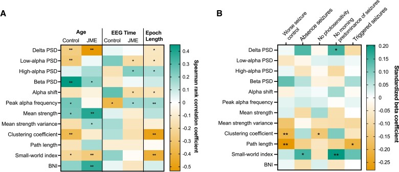
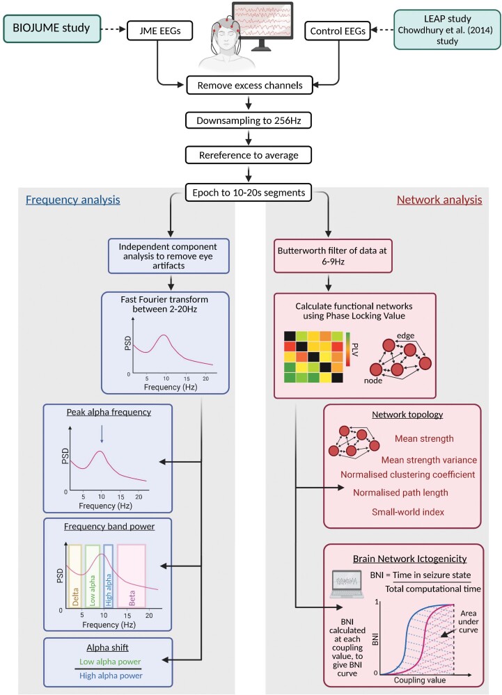
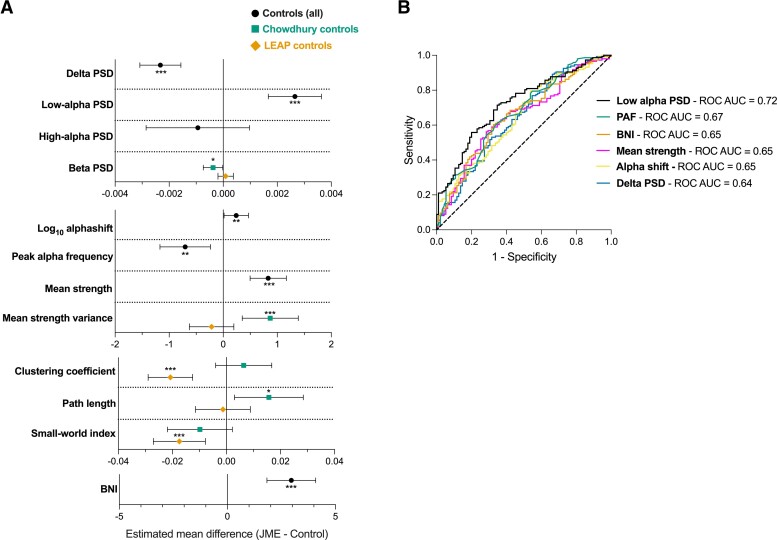
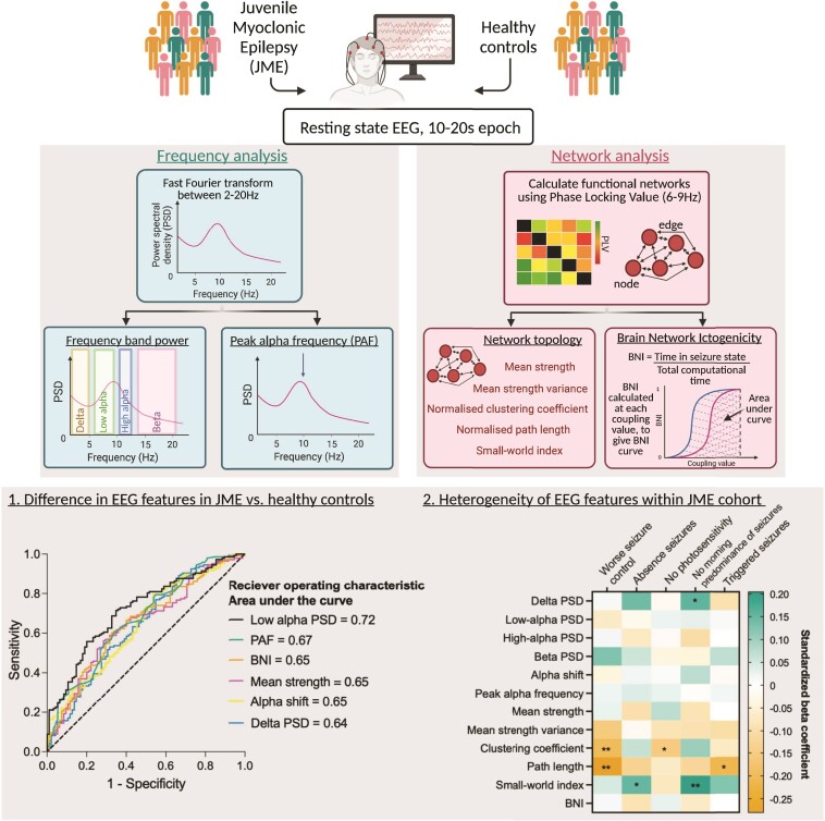
Abnormal EEG features are a hallmark of epilepsy, and abnormal frequency and network features are apparent in EEGs from people with idiopathic generalized epilepsy in both ictal and interictal states. Here, we characterize differences in the resting-state EEG of individuals with juvenile myoclonic epilepsy and assess factors influencing the heterogeneity of EEG features. We collected EEG data from 147 participants with juvenile myoclonic epilepsy through the Biology of Juvenile Myoclonic Epilepsy study. Ninety-five control EEGs were acquired from two independent studies [Chowdhury . (2014) and EU-AIMS Longitudinal European Autism Project]. We extracted frequency and functional network-based features from 10 to 20 s epochs of resting-state EEG, including relative power spectral density, peak alpha frequency, network topology measures and brain network ictogenicity: a computational measure of the propensity of networks to generate seizure dynamics. We tested for differences between epilepsy and control EEGs using univariate, multivariable and receiver operating curve analysis. In addition, we explored the heterogeneity of EEG features within and between cohorts by testing for associations with potentially influential factors such as age, sex, epoch length and time, as well as testing for associations with clinical phenotypes including anti-seizure medication, and seizure characteristics in the epilepsy cohort. -values were corrected for multiple comparisons. Univariate analysis showed significant differences in power spectral density in delta (2-5 Hz) ( = 0.0007, hedges' g = 0.55) and low-alpha (6-9 Hz) ( = 2.9 × 10, g = 0.80) frequency bands, peak alpha frequency ( = 0.000007, g = 0.66), functional network mean degree ( = 0.0006, g = 0.48) and brain network ictogenicity ( = 0.00006, g = 0.56) between epilepsy and controls. Since age ( = 0.009) and epoch length ( = 1.7 × 10) differed between the two groups and were potential confounders, we controlled for these covariates in multivariable analysis where disparities in EEG features between epilepsy and controls remained. Receiver operating curve analysis showed low-alpha power spectral density was optimal at distinguishing epilepsy from controls, with an area under the curve of 0.72. Lower average normalized clustering coefficient and shorter average normalized path length were associated with poorer seizure control in epilepsy patients. To conclude, individuals with juvenile myoclonic epilepsy have increased power of neural oscillatory activity at low-alpha frequencies, and increased brain network ictogenicity compared with controls, supporting evidence from studies in other epilepsies with considerable external validity. In addition, the impact of confounders on different frequency-based and network-based EEG features observed in this study highlights the need for careful consideration and control of these factors in future EEG research in idiopathic generalized epilepsy particularly for their use as biomarkers.
异常脑电图特征是癫痫的一个标志,在特发性全身性癫痫患者的发作期和发作间期脑电图中,异常频率和网络特征都很明显。在此,我们描述青少年肌阵挛性癫痫患者静息态脑电图的差异,并评估影响脑电图特征异质性的因素。我们通过青少年肌阵挛性癫痫生物学研究,收集了147名青少年肌阵挛性癫痫患者的脑电图数据。从两项独立研究[乔杜里(2014年)和欧盟-目标纵向欧洲自闭症项目]中获取了95份对照脑电图。我们从静息态脑电图的10至20秒时段中提取了基于频率和功能网络的特征,包括相对功率谱密度、峰值阿尔法频率、网络拓扑测量和脑网络致痫性:一种衡量网络产生癫痫发作动态倾向的计算指标。我们使用单变量、多变量和受试者工作特征曲线分析来测试癫痫脑电图与对照脑电图之间的差异。此外,我们通过测试与年龄、性别、时段长度和时间等潜在影响因素的关联,以及与癫痫队列中的抗癫痫药物和发作特征等临床表型的关联,来探索队列内部和队列之间脑电图特征的异质性。对p值进行了多重比较校正。单变量分析显示,在δ(2 - 5赫兹)(p = 0.0007,赫奇斯效应量g = 0.55)和低阿尔法(6 - 9赫兹)(p = 2.9×10⁻⁵,g = 0.80)频段的功率谱密度、峰值阿尔法频率(p = 0.000007,g = 0.66)、功能网络平均度(p = 0.0006,g = 0.48)和脑网络致痫性(p = 0.00006,g = 0.56)方面,癫痫患者与对照组之间存在显著差异。由于两组之间年龄(p = 0.009)和时段长度(p = 1.7×10⁻⁴)不同且是潜在混杂因素,我们在多变量分析中对这些协变量进行了控制,而癫痫患者与对照组之间脑电图特征的差异仍然存在。受试者工作特征曲线分析显示,低阿尔法功率谱密度在区分癫痫与对照组方面最为理想,曲线下面积为0.72。癫痫患者中较低的平均归一化聚类系数和较短的平均归一化路径长度与较差的发作控制相关。总之,与对照组相比,青少年肌阵挛性癫痫患者在低阿尔法频率下神经振荡活动的功率增加,脑网络致痫性增加,这支持了其他癫痫研究中具有相当外部效度的证据。此外,本研究中观察到的混杂因素对不同基于频率和基于网络的脑电图特征的影响,凸显了在未来特发性全身性癫痫的脑电图研究中,特别是将其用作生物标志物时,需要仔细考虑和控制这些因素。