Department of Ophthalmology, Samsung Medical Center, Sungkyunkwan University School of Medicine, 81 Irwon-ro, Gangnam-gu, Seoul, 06351, Republic of Korea.
Department of Neurology, Samsung Medical Center, Sungkyunkwan University School of Medicine, 81 Irwon-ro, Gangnam-gu, Seoul, 06351, Republic of Korea.
Alzheimers Res Ther. 2022 Jul 25;14(1):99. doi: 10.1186/s13195-022-01045-0.
Decreased visual acuity (VA) is reported to be a risk factor for dementia. However, the association between VA and cortical thickness has not been established. We investigated the association between VA and cortical thickness in cognitively normal adults.
We conducted a cross-sectional, single-center cohort study with cognitively normal adults (aged ≥ 45) who received medical screening examinations at the Health Promotion Center at Samsung Medical Center. Subjects were categorized as bad (VA ≤ 20/40), fair (20/40 < VA ≤ 20/25), and good (VA > 20/25) VA group by using corrected VA in the Snellen system. Using 3D volumetric brain MRI, cortical thickness was calculated using the Euclidean distance between the linked vertices of the inner and outer surfaces. We analyzed the association between VA and cortical thickness after controlling for age, sex, hypertension, diabetes, dyslipidemia, intracranial volume, and education level.
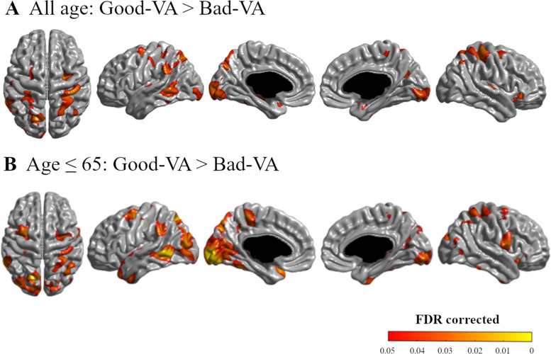
A total of 2756 subjects were analyzed in this study. Compared to the good VA group, the bad VA group showed overall thinner cortex (p = 0.015), especially in the parietal (p = 0.018) and occipital (p = 0.011) lobes. Topographical color maps of vertex-wise analysis also showed that the bad VA group showed a thinner cortex in the parieto-temporo-occipital area. These results were more robust in younger adults (aged 45 to 65) as decreased VA was associated with thinner cortex in more widespread regions in the parieto-temporo-occipital area.
Our results suggest that a thinner cortex in the visual processing area of the brain is related to decreased visual stimuli.
视力下降(VA)被报道为痴呆的危险因素。然而,VA 与皮质厚度之间的关联尚未确定。我们研究了认知正常成年人中 VA 与皮质厚度之间的关系。
我们进行了一项横断面、单中心队列研究,纳入了在三星医疗中心健康促进中心接受医疗筛查的认知正常成年人(年龄≥45 岁)。通过 Snellen 系统中的矫正视力,将受试者分为视力差(VA≤20/40)、视力一般(20/40<VA≤20/25)和视力良好(VA>20/25)组。使用 3D 容积脑 MRI,通过内、外表面的连接顶点之间的欧几里得距离计算皮质厚度。我们在控制年龄、性别、高血压、糖尿病、血脂异常、颅内体积和教育程度后,分析了 VA 与皮质厚度之间的关系。
本研究共分析了 2756 名受试者。与视力良好组相比,视力差组的皮质整体较薄(p=0.015),尤其是顶叶(p=0.018)和枕叶(p=0.011)。顶点分析的拓扑彩色图谱还显示,视力差组在顶颞枕叶区域的皮质较薄。这些结果在较年轻的成年人(45 至 65 岁)中更为可靠,因为 VA 降低与顶颞枕叶区域更广泛的区域的皮质变薄有关。
我们的结果表明,大脑视觉处理区域的皮质变薄与视觉刺激减少有关。