Dong Xiuming, Tse Gary, Hao Guoliang, Du Yimei
Henan SCOPE Research Institute of Electrophysiology Co., Ltd., Kaifeng 475000, China.
Cardiac Electrophysiology Unit, Cardiovascular Analytics Group, Hong Kong, China.
Life (Basel). 2022 Jul 5;12(7):996. doi: 10.3390/life12070996.
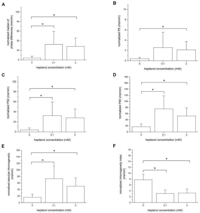
Background: Previous studies have associated slowed ventricular conduction with the arrhythmogenesis mediated by the gap junction and sodium channel inhibitor heptanol in mouse hearts. However, they did not study the propagation patterns that might contribute to the arrhythmic substrate. This study used a multi-electrode array mapping technique to further investigate different conduction abnormalities in Langendorff-perfused mouse hearts exposed to 0.1 or 2 mM heptanol. Methods: Recordings were made from the left ventricular epicardium using multi-electrode arrays in spontaneously beating hearts during right ventricular 8 Hz pacing or S1S2 pacing. Results: In spontaneously beating hearts, heptanol at 0.1 and 2 mM significantly reduced the heart rate from 314 ± 25 to 189 ± 24 and 157 ± 7 bpm, respectively (ANOVA, p < 0.05 and p < 0.001). During regular 8 Hz pacing, the mean LATs were increased by 0.1 and 2 mM heptanol from 7.1 ± 2.2 ms to 19.9 ± 5.0 ms (p < 0.05) and 18.4 ± 5.7 ms (p < 0.05). The standard deviation of the mean LATs was increased from 2.5 ± 0.8 ms to 10.3 ± 4.0 ms and 8.0 ± 2.5 ms (p < 0.05), and the median of phase differences was increased from 1.7 ± 1.1 ms to 13.9 ± 7.8 ms and 12.1 ± 5.0 ms by 0.1 and 2 mM heptanol (p < 0.05). P5 took a value of 0.2 ± 0.1 ms and was not significantly altered by heptanol at 0.1 or 2 mM (1.1 ± 0.9 ms and 0.9 ± 0.5 ms, p > 0.05). P50 was increased from 7.3 ± 2.7 ms to 24.0 ± 12.0 ms by 0.1 mM heptanol and then to 22.5 ± 7.5 ms by 2 mM heptanol (p < 0.05). P95 was increased from 1.7 ± 1.1 ms to 13.9 ± 7.8 ms by 0.1 mM heptanol and to 12.1 ± 5.0 ms by 2 mM heptanol (p < 0.05). These changes led to increases in the absolute inhomogeneity in conduction (P5−95) from 7.1 ± 2.6 ms to 31.4 ± 11.3 ms, 2 mM: 21.6 ± 7.2 ms, respectively (p < 0.05). The inhomogeneity index (P5−95/P50) was significantly reduced from 3.7 ± 1.2 to 3.1 ± 0.8 by 0.1 mM and then to 3.3 ± 0.9 by 2 mM heptanol (p < 0.05). Conclusion: Increased activation latencies, reduced CVs, and the increased inhomogeneity index of conduction were associated with both spontaneous and induced ventricular arrhythmias.
先前的研究已将心室传导减慢与缝隙连接和钠通道抑制剂庚醇介导的小鼠心脏心律失常发生联系起来。然而,他们并未研究可能导致心律失常基质的传导模式。本研究使用多电极阵列映射技术,进一步研究暴露于0.1或2 mM庚醇的Langendorff灌注小鼠心脏中的不同传导异常。方法:在右心室8 Hz起搏或S1S2起搏期间,使用多电极阵列从自发跳动心脏的左心室心外膜进行记录。结果:在自发跳动的心脏中,0.1和2 mM的庚醇分别使心率从314±25显著降低至189±24和157±7次/分钟(方差分析,p < 0.05和p < 0.001)。在规则的8 Hz起搏期间,0.1和2 mM庚醇使平均局部激活时间(LATs)从7.1±2.2 ms增加至19.9±5.0 ms(p < 0.05)和18.4±5.7 ms(p < 0.05)。平均LATs的标准差从2.5±0.8 ms增加至10.3±4.0 ms和8.0±2.5 ms(p < 0.05),0.1和2 mM庚醇使相差中位数从1.7±1.1 ms增加至13.9±7.8 ms和12.1±5.0 ms(p < 0.05)。P5值为0.2±0.1 ms,0.1或2 mM庚醇对其无显著改变(分别为1.1±0.9 ms和0.9±0.5 ms,p > 0.05)。0.1 mM庚醇使P50从7.3±2.7 ms增加至24.0±12.0 ms,然后2 mM庚醇使其增加至22.5±7.5 ms(p < 0.05)。0.1 mM庚醇使P95从1.7±1.1 ms增加至13.9±7.8 ms,2 mM庚醇使其增加至12.1±5.0 ms(p < 0.05)。这些变化导致传导的绝对不均匀性(P5−95)分别从7.1±2.6 ms增加至31.4±11.3 ms、2 mM时为21.6±7.2 ms(p < 0.05)。不均匀性指数(P5−95/P50)在0.1 mM时从3.7±1.2显著降低至3.1±0.8,然后2 mM庚醇使其降至3.3±0.9(p < 0.05)。结论:激活延迟增加、传导速度降低以及传导不均匀性指数增加与自发性和诱发性室性心律失常相关。