• 文献检索
  • 文档翻译
  • 深度研究
  • 学术资讯
  • Suppr Zotero 插件Zotero 插件
  • 邀请有礼
  • 套餐&价格
  • 历史记录
应用&插件
Suppr Zotero 插件Zotero 插件浏览器插件Mac 客户端Windows 客户端微信小程序
定价
高级版会员购买积分包购买API积分包
服务
文献检索文档翻译深度研究API 文档MCP 服务
关于我们
关于 Suppr公司介绍联系我们用户协议隐私条款
关注我们

Suppr 超能文献

核心技术专利:CN118964589B侵权必究
粤ICP备2023148730 号-1Suppr @ 2026

文献检索

告别复杂PubMed语法,用中文像聊天一样搜索,搜遍4000万医学文献。AI智能推荐,让科研检索更轻松。

立即免费搜索

文件翻译

保留排版,准确专业,支持PDF/Word/PPT等文件格式,支持 12+语言互译。

免费翻译文档

深度研究

AI帮你快速写综述,25分钟生成高质量综述,智能提取关键信息,辅助科研写作。

立即免费体验

一种基于复杂网络的肺部图像处理新方法

A Novel Method for Lung Image Processing Using Complex Networks.

机构信息

Department of Computer and Information Technology, Automation and Computers Faculty, "Politehnica" University of Timișoara, Vasile Pârvan Blvd. No. 2, 300223 Timișoara, Romania.

Pulmonology Department, 'Victor Babes' University of Medicine and Pharmacy, Eftimie Murgu Square 2, 300041 Timișoara, Romania.

出版信息

Tomography. 2022 Jul 27;8(4):1928-1946. doi: 10.3390/tomography8040162.

DOI:10.3390/tomography8040162
PMID:35894027
原文链接:https://pmc.ncbi.nlm.nih.gov/articles/PMC9332806/
Abstract

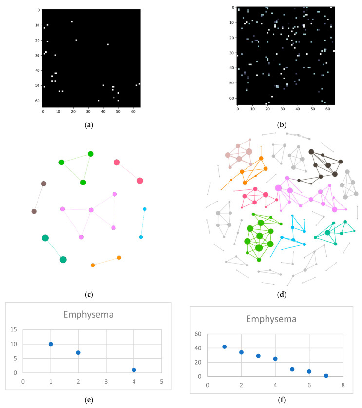
The High-Resolution Computed Tomography (HRCT) detection and diagnosis of diffuse lung disease is primarily based on the recognition of a limited number of specific abnormal findings, pattern combinations or their distributions, as well as anamnesis and clinical information. Since texture recognition has a very high accuracy percentage if a complex network approach is used, this paper aims to implement such a technique customized for diffuse interstitial lung diseases (DILD). The proposed procedure translates HRCT lung imaging into complex networks by taking samples containing a secondary lobule, converting them into complex networks and analyzing them in three dimensions: emphysema, ground glass opacity, and consolidation. This method was evaluated on a 60-patient lot and the results showed a clear, quantifiable difference between healthy and affected lungs. By deconstructing the image on three pathological axes, the method offers an objective way to quantify DILD details which, so far, have only been analyzed subjectively.

摘要

高分辨率计算机断层扫描(HRCT)对弥漫性肺疾病的检测和诊断主要基于对有限数量的特定异常发现、模式组合或其分布的识别,以及病史和临床信息。由于纹理识别在使用复杂网络方法时具有非常高的准确率百分比,因此本文旨在为弥漫性间质性肺病(DILD)实现这种技术。所提出的方法通过对包含次级小叶的样本进行处理,将 HRCT 肺部成像转换为复杂网络,并在三个维度上对其进行分析:肺气肿、磨玻璃影和实变。该方法在 60 名患者的样本上进行了评估,结果表明健康和患病肺部之间存在明显的、可量化的差异。通过对三个病理轴进行图像分解,该方法提供了一种客观的方法来量化 DILD 细节,这些细节迄今为止仅进行了主观分析。

https://cdn.ncbi.nlm.nih.gov/pmc/blobs/d609/9332806/132b7afb73eb/tomography-08-00162-g013.jpg
https://cdn.ncbi.nlm.nih.gov/pmc/blobs/d609/9332806/db0931969963/tomography-08-00162-g001.jpg
https://cdn.ncbi.nlm.nih.gov/pmc/blobs/d609/9332806/9a47ce05f24a/tomography-08-00162-g002.jpg
https://cdn.ncbi.nlm.nih.gov/pmc/blobs/d609/9332806/3c5684e73297/tomography-08-00162-g003.jpg
https://cdn.ncbi.nlm.nih.gov/pmc/blobs/d609/9332806/b323c98856f9/tomography-08-00162-g004.jpg
https://cdn.ncbi.nlm.nih.gov/pmc/blobs/d609/9332806/41e05558b9e4/tomography-08-00162-g005.jpg
https://cdn.ncbi.nlm.nih.gov/pmc/blobs/d609/9332806/abdcc6d0b9d2/tomography-08-00162-g006.jpg
https://cdn.ncbi.nlm.nih.gov/pmc/blobs/d609/9332806/eddd436958dd/tomography-08-00162-g007.jpg
https://cdn.ncbi.nlm.nih.gov/pmc/blobs/d609/9332806/c3a389e12358/tomography-08-00162-g008.jpg
https://cdn.ncbi.nlm.nih.gov/pmc/blobs/d609/9332806/27f1a37f58f2/tomography-08-00162-g009.jpg
https://cdn.ncbi.nlm.nih.gov/pmc/blobs/d609/9332806/37582452349a/tomography-08-00162-g010.jpg
https://cdn.ncbi.nlm.nih.gov/pmc/blobs/d609/9332806/8c7a1f1d5c78/tomography-08-00162-g011.jpg
https://cdn.ncbi.nlm.nih.gov/pmc/blobs/d609/9332806/93be4ee14dd6/tomography-08-00162-g012.jpg
https://cdn.ncbi.nlm.nih.gov/pmc/blobs/d609/9332806/132b7afb73eb/tomography-08-00162-g013.jpg

相似文献

1
A Novel Method for Lung Image Processing Using Complex Networks.一种基于复杂网络的肺部图像处理新方法
Tomography. 2022 Jul 27;8(4):1928-1946. doi: 10.3390/tomography8040162.
2
Enhancing Imagistic Interstitial Lung Disease Diagnosis by Using Complex Networks.利用复杂网络增强间质性肺病的影像学诊断
Medicina (Kaunas). 2022 Sep 16;58(9):1288. doi: 10.3390/medicina58091288.
3
Lung Segmentation on HRCT and Volumetric CT for Diffuse Interstitial Lung Disease Using Deep Convolutional Neural Networks.基于深度卷积神经网络的 HRCT 和容积 CT 对弥漫性间质性肺疾病的肺分割。
J Digit Imaging. 2019 Dec;32(6):1019-1026. doi: 10.1007/s10278-019-00254-8.
4
Recent advances in radiology of the interstitial lung disease.间质性肺疾病的放射学最新进展。
Curr Opin Pulm Med. 1998 Sep;4(5):281-7. doi: 10.1097/00063198-199809000-00007.
5
The diagnostic accuracy of high-resolution computed tomography in diffuse infiltrative lung diseases.高分辨率计算机断层扫描在弥漫性浸润性肺疾病中的诊断准确性。
Chest. 1993 Oct;104(4):1149-55. doi: 10.1378/chest.104.4.1149.
6
Perfusion- and pattern-based quantitative CT indexes using contrast-enhanced dual-energy computed tomography in diffuse interstitial lung disease: relationships with physiologic impairment and prediction of prognosis.在弥漫性间质性肺疾病中使用对比增强双能量计算机断层扫描的基于灌注和模式的定量CT指标:与生理功能损害的关系及预后预测
Eur Radiol. 2016 May;26(5):1368-77. doi: 10.1007/s00330-015-3946-2. Epub 2015 Aug 9.
7
Feasibility of automated quantification of regional disease patterns depicted on high-resolution computed tomography in patients with various diffuse lung diseases.对各种弥漫性肺部疾病患者的高分辨率计算机断层扫描所显示的区域疾病模式进行自动定量分析的可行性。
Korean J Radiol. 2009 Sep-Oct;10(5):455-63. doi: 10.3348/kjr.2009.10.5.455. Epub 2009 Aug 25.
8
Radiological evaluation of interstitial lung disease.间质性肺疾病的放射学评估。
Curr Opin Pulm Med. 2011 Sep;17(5):337-45. doi: 10.1097/MCP.0b013e328347c16a.
9
High-resolution CT in diagnosis of diffuse infiltrative lung disease.高分辨率CT在弥漫性浸润性肺疾病诊断中的应用
Semin Ultrasound CT MR. 2005 Oct;26(5):332-47. doi: 10.1053/j.sult.2005.07.004.
10
High-resolution computed tomography patterns of diffuse interstitial lung disease with clinical and pathological correlation.弥漫性间质性肺疾病的高分辨率计算机断层扫描模式及其临床与病理相关性
J Bras Pneumol. 2008 Sep;34(9):715-44. doi: 10.1590/s1806-37132008000900013.

引用本文的文献

1
Enhancing Adult Asthma Management: A Review on the Utility of Remote Home Spirometry and Mobile Applications.加强成人哮喘管理:关于远程家庭肺功能测定和移动应用程序效用的综述
J Pers Med. 2024 Aug 11;14(8):852. doi: 10.3390/jpm14080852.
2
Stratifying Disease Severity in Pediatric COVID-19: A Correlative Study of Serum Biomarkers and Lung Ultrasound-A Retrospective Observational Dual-Center Study.儿童新冠病毒病的疾病严重程度分层:血清生物标志物与肺部超声的相关性研究——一项回顾性观察性双中心研究
Diagnostics (Basel). 2024 Feb 17;14(4):440. doi: 10.3390/diagnostics14040440.
3
The Impact of Therapeutic Plasma Exchange on Inflammatory Markers and Acute Phase Reactants in Patients with Severe SARS-CoV-2 Infection.

本文引用的文献

1
Interstitial Lung Disease in 2020: A History of Progress.2020 年间质性肺病:进展历程。
Clin Chest Med. 2021 Jun;42(2):229-239. doi: 10.1016/j.ccm.2021.03.001.
2
Practical Imaging Interpretation in Patients Suspected of Having Idiopathic Pulmonary Fibrosis: Official Recommendations from the Radiology Working Group of the Pulmonary Fibrosis Foundation.疑似特发性肺纤维化患者的实用影像解读:肺纤维化基金会放射学工作组的官方建议
Radiol Cardiothorac Imaging. 2021 Feb 25;3(1):e200279. doi: 10.1148/ryct.2021200279. eCollection 2021 Feb.
3
Explaining the emergence of complex networks through log-normal fitness in a Euclidean node similarity space.
治疗性血浆置换对重症 SARS-CoV-2 感染患者炎症标志物和急性期反应物的影响。
Medicina (Kaunas). 2023 Apr 29;59(5):867. doi: 10.3390/medicina59050867.
4
Laboratory Findings and Clinical Outcomes of ICU-admitted COVID-19 Patients: A Retrospective Assessment of Particularities Identified among Romanian Minorities.入住重症监护病房的新冠病毒肺炎患者的实验室检查结果及临床结局:对罗马尼亚少数民族中发现的特殊性进行的回顾性评估
J Pers Med. 2023 Jan 21;13(2):195. doi: 10.3390/jpm13020195.
5
A Retrospective Assessment of Laboratory Findings and Cytokine Markers in Severe SARS-CoV-2 Infection among Patients of Roma Population.罗姆人群中重症新型冠状病毒2型感染患者实验室检查结果及细胞因子标志物的回顾性评估
J Clin Med. 2022 Nov 16;11(22):6777. doi: 10.3390/jcm11226777.
6
Disease Progression, Clinical Features, and Risk Factors for Pneumonia in Unvaccinated Children and Adolescents with Measles: A Re-Emerging Disease in Romania.未接种麻疹疫苗的儿童和青少年麻疹患者的疾病进展、临床特征和肺炎风险因素:罗马尼亚再现的疾病。
Int J Environ Res Public Health. 2022 Oct 13;19(20):13165. doi: 10.3390/ijerph192013165.
7
Enhancing Imagistic Interstitial Lung Disease Diagnosis by Using Complex Networks.利用复杂网络增强间质性肺病的影像学诊断
Medicina (Kaunas). 2022 Sep 16;58(9):1288. doi: 10.3390/medicina58091288.
通过欧几里得节点相似空间中的对数正态适应度解释复杂网络的出现。
Sci Rep. 2021 Jan 21;11(1):1976. doi: 10.1038/s41598-021-81547-3.
4
Automated CT Analysis of Major Forms of Interstitial Lung Disease.间质性肺疾病主要类型的CT自动分析
J Clin Med. 2020 Nov 23;9(11):3776. doi: 10.3390/jcm9113776.
5
COVID-19 pneumonia: computer-aided quantification of healthy lung parenchyma, emphysema, ground glass and consolidation on chest computed tomography (CT).COVID-19 肺炎:胸部计算机断层扫描(CT)上健康肺实质、肺气肿、磨玻璃影和实变的计算机辅助定量。
Radiol Med. 2021 Apr;126(4):553-560. doi: 10.1007/s11547-020-01305-9. Epub 2020 Nov 18.
6
Clinical impact of the radiological indeterminate for usual interstitial pneumonia pattern on the diagnosis of idiopathic pulmonary fibrosis.影像学不确定的寻常型间质性肺炎模式对特发性肺纤维化诊断的临床影响。
Respir Investig. 2021 Jan;59(1):81-89. doi: 10.1016/j.resinv.2020.07.001. Epub 2020 Aug 28.
7
Deep learning in interstitial lung disease-how long until daily practice.深度学习在间质性肺疾病中的应用——离日常实践还有多久。
Eur Radiol. 2020 Nov;30(11):6285-6292. doi: 10.1007/s00330-020-06986-4. Epub 2020 Jun 14.
8
Artificial intelligence to codify lung CT in Covid-19 patients.人工智能对新冠病毒患者肺部 CT 进行编码。
Radiol Med. 2020 May;125(5):500-504. doi: 10.1007/s11547-020-01195-x. Epub 2020 May 4.
9
Using Artificial Intelligence to Detect COVID-19 and Community-acquired Pneumonia Based on Pulmonary CT: Evaluation of the Diagnostic Accuracy.基于肺部 CT 的人工智能检测 COVID-19 和社区获得性肺炎:诊断准确性评估。
Radiology. 2020 Aug;296(2):E65-E71. doi: 10.1148/radiol.2020200905. Epub 2020 Mar 19.
10
Quantitative CT Analysis of Diffuse Lung Disease.弥漫性肺疾病的定量 CT 分析。
Radiographics. 2020 Jan-Feb;40(1):28-43. doi: 10.1148/rg.2020190099. Epub 2019 Nov 29.