Department of Biomedical Imaging and Image-Guided Therapy, General Hospital of Vienna (AKH), Medical University of Vienna, Waehringer Guertel 18-20, 1090, Vienna, Austria.
Eur Radiol. 2023 Jan;33(1):523-534. doi: 10.1007/s00330-022-08984-0. Epub 2022 Jul 27.
To investigate the effect of saline-diluted gadoxetic acid, done for arterial-phase (AP) artifact reduction, on signal intensity (SI), and hence focal lesion conspicuity on MR imaging.
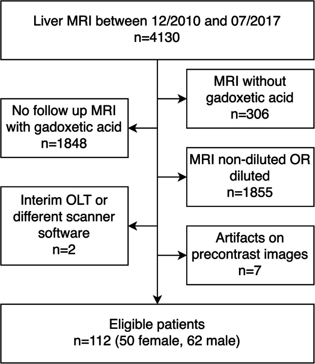
We retrospectively examined 112 patients who each had at least two serial gadoxetic acid-enhanced liver MRIs performed at 1 ml/s, first with non-diluted (ND), then with 1:1 saline-diluted (D) contrast. Two blinded readers independently analyzed the artifacts and graded dynamic images using a 5-point scale. The absolute SI of liver parenchyma, focal liver lesions (if present), aorta, and portal vein at the level of the celiac trunk and the SI of the paraspinal muscle were measured in all phases. The signal-to-norm (SI) of the vascular structures, hepatic parenchyma and focal lesions, and the contrast-to-norm (C) of focal liver lesions were calculated.
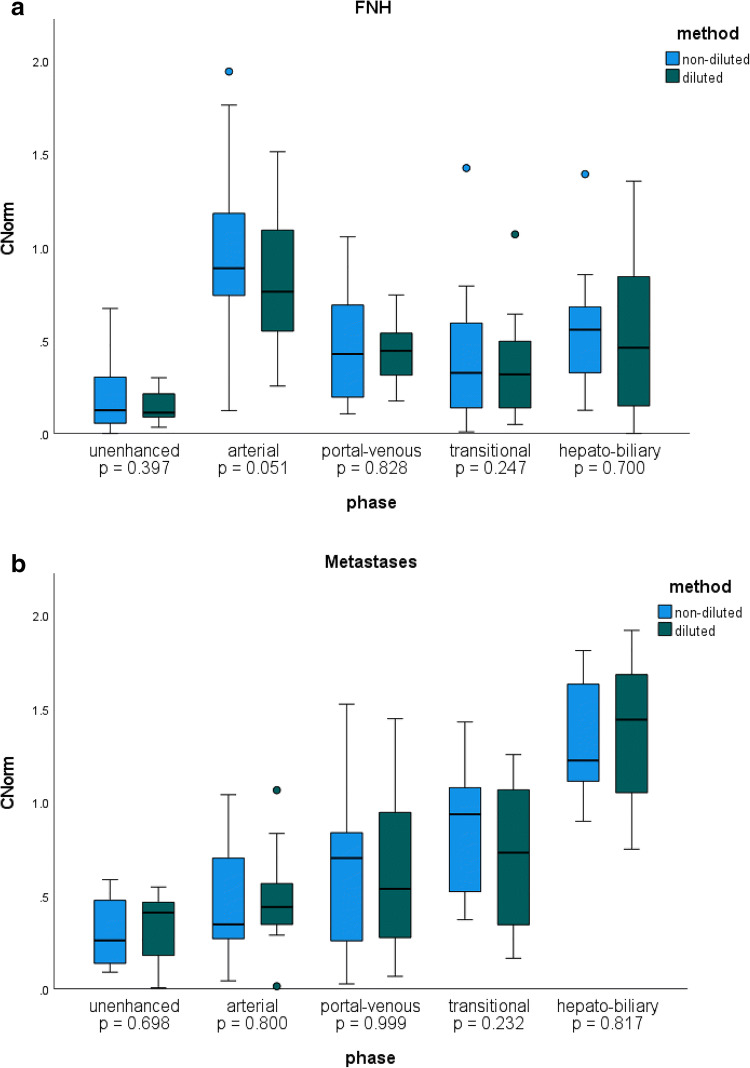
AP artifacts were significantly reduced with dilution. Mean absolute contrast-enhanced liver SI was significantly higher on the D exams compared to the ND exams. Likewise, SI of liver parenchyma was significantly higher in all contrast-enhanced phases except transitional phase on the D exams. SI values in the AP for the aorta and in the PVP for portal vein were significantly higher on the diluted exams. The C was not significantly different between ND and D exams for lesions in any imaging phase. The interclass correlation coefficient was excellent (0.89).
Gadoxetic acid dilution injected at 1ml/s produces images with significantly fewer AP artifacts but no significant loss in SI or C compared to standard non-diluted images.
• Diluted gadoxetic acid at slow injection (1 ml/s) yielded images with higher SI of the liver parenchyma and preserved C for focal liver lesions. • Gadoxetic acid-enhanced MRI injected at 1 ml/s is associated with arterial-phase (AP) artifacts in 31% of exams, which may degrade image quality and limits focal liver lesion detection. • Saline dilution of gadoxetic acid 1:1 combined with a slow injection rate of 1 ml/s significantly reduced AP artifacts from 31 to 9% and non-diagnostic AP artifacts from 16 to 1%.
探讨用于动脉期(AP)伪影减少的盐水稀释钆塞酸,对磁共振成像(MRI)信号强度(SI)的影响,进而对局灶性病变的显影效果。
我们回顾性地检查了 112 名患者,他们每个人都进行了至少两次以 1ml/s 速度进行的钆塞酸增强肝脏 MRI 检查,第一次使用非稀释(ND),然后使用 1:1 盐水稀释(D)对比剂。两名盲法读者使用 5 分制独立分析伪影,并对动态图像进行分级。在所有阶段测量腹腔干水平的肝实质、局灶性肝病变(如有)、主动脉和门静脉的绝对 SI,以及椎旁肌肉的 SI。测量血管结构、肝实质和局灶性病变的信号与正常(SI)比,以及局灶性肝病变的对比与正常(C)比。
稀释后 AP 伪影显著减少。与 ND 检查相比,D 检查的肝实质平均增强 SI 显著升高。同样,除过渡相外,D 检查的所有增强相的肝实质 SI 均升高。稀释后,AP 中的主动脉和 PVP 中的门静脉的 SI 值升高。在任何成像阶段,病变的 C 值在 ND 和 D 检查之间均无显著差异。ICC 为 0.89。
以 1ml/s 注入的钆塞酸稀释液可产生 AP 伪影显著减少的图像,但与标准非稀释图像相比,SI 或 C 无显著损失。
• 以缓慢(1ml/s)注射的稀释钆塞酸可产生肝实质 SI 更高、局灶性肝病变的 C 值保持不变的图像。
• 以 1ml/s 速度注入的钆塞酸增强 MRI 在 31%的检查中与动脉期(AP)伪影相关,这可能会降低图像质量,限制局灶性肝病变的检测。
• 1:1 盐水稀释的钆塞酸与 1ml/s 的缓慢注射速率相结合,可将 AP 伪影从 31%显著减少至 9%,并将无诊断价值的 AP 伪影从 16%减少至 1%。