Department of Experimental and Clinical Biomedical Sciences "Mario Serio", University of Florence, 50134 Florence, Italy.
Department of Experimental Medicine, Cellular Electron Microscopy Laboratory, University of Genova, 16132 Genova, Italy.
Sci Adv. 2022 Jul 29;8(30):eabm6376. doi: 10.1126/sciadv.abm6376. Epub 2022 Jul 27.
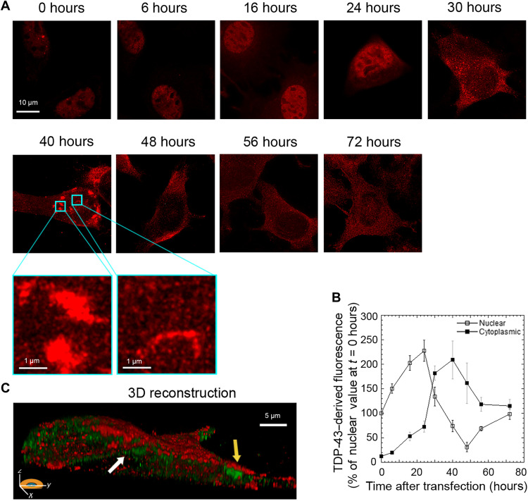
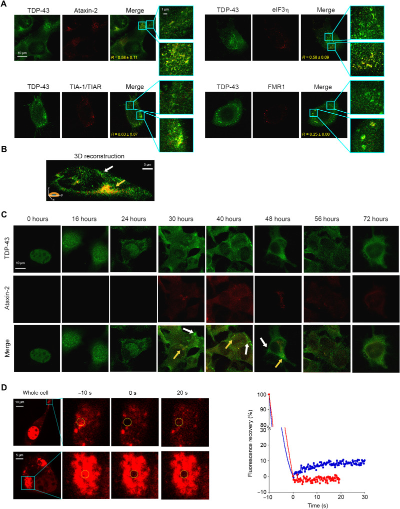
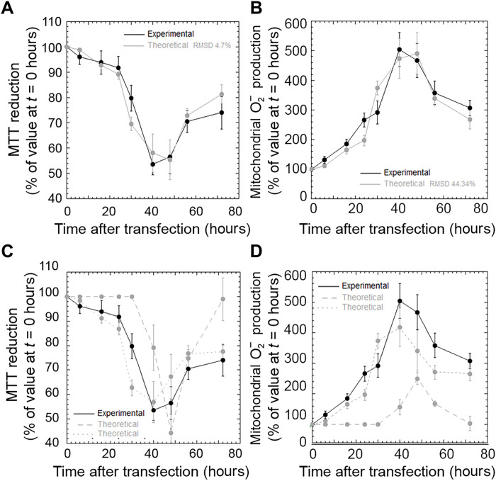
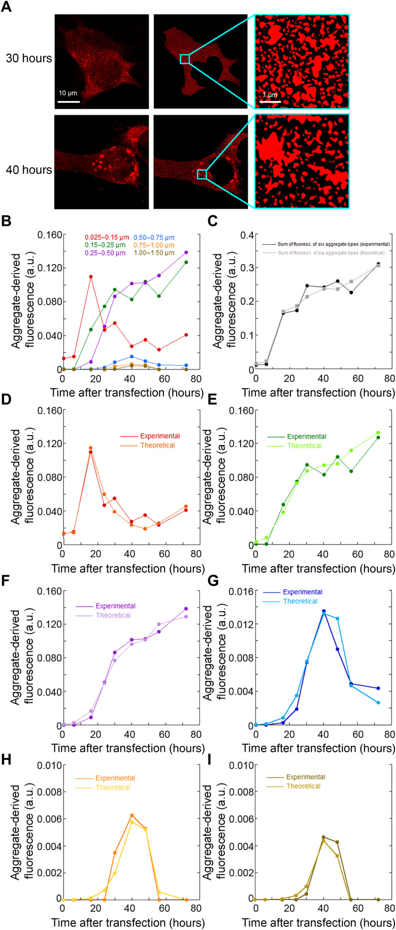
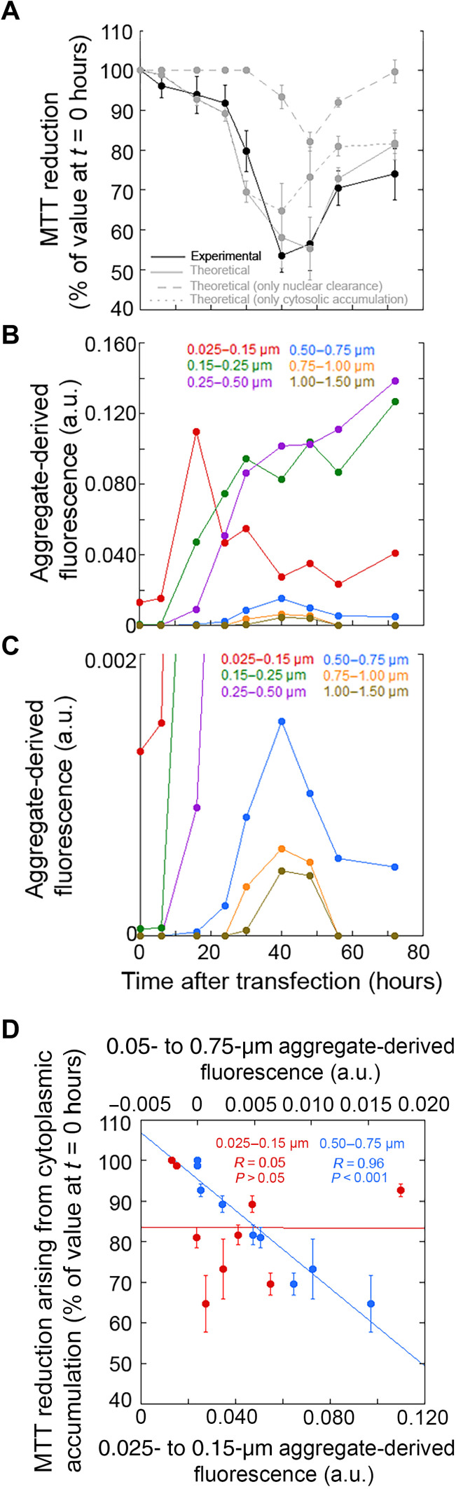
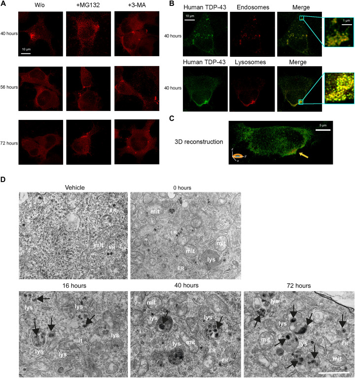
A number of neurodegenerative conditions are associated with the formation of cytosolic inclusions of TDP-43 within neurons. We expressed full-length TDP-43 in a motoneuron/neuroblastoma hybrid cell line (NSC-34) and exploited the high-resolution power of stimulated emission depletion microscopy to monitor the changes of nuclear and cytoplasmic TDP-43 levels and the formation of various size classes of cytoplasmic TDP-43 aggregates with time. Concomitantly, we monitored oxidative stress and mitochondrial impairment using the MitoSOX and MTT reduction assays, respectively. Using a quantitative biology approach, we attributed neuronal dysfunction associated with cytoplasmic deposition component to the formation of the largest inclusions, independently of stress granules. This is in contrast to other neurodegenerative diseases where toxicity is attributed to small oligomers. Using specific inhibitors, markers, and electron microscopy, the proteasome and autophagy were found to target mainly the largest deleterious inclusions, but their efficiency soon decreases without full recovery of neuronal viability.
许多神经退行性疾病与神经元细胞浆中 TDP-43 的形成有关。我们在运动神经元/神经母细胞瘤杂交细胞系(NSC-34)中表达全长 TDP-43,并利用受激发射损耗显微镜的高分辨率能力来监测核和细胞质 TDP-43 水平的变化以及随时间推移形成的各种大小类别的细胞质 TDP-43 聚集体。同时,我们分别使用 MitoSOX 和 MTT 还原测定法监测氧化应激和线粒体损伤。使用定量生物学方法,我们将与细胞质沉积成分相关的神经元功能障碍归因于最大聚集体的形成,而与应激颗粒无关。这与其他神经退行性疾病形成对比,在其他神经退行性疾病中,毒性归因于小的寡聚物。使用特定的抑制剂、标记物和电子显微镜,发现蛋白酶体和自噬主要靶向最大的有害聚集体,但它们的效率很快下降,而神经元活力没有完全恢复。