Department of Anatomical Sciences, Shiraz Medical School, Shiraz University of Medical Sciences, Shiraz, Iran.
Histomorphometry and Stereology Research Center, Shiraz Medical School, Shiraz University of Medical Sciences, Shiraz, Iran.
Vet Med Sci. 2022 Sep;8(5):2092-2103. doi: 10.1002/vms3.891. Epub 2022 Jul 27.
To prevent ovarian hyperstimulation syndrome, in vitro maturation (IVM) allows the oocytes for infertility treatment without hormone therapy. Although many oocytes matured during IVM, some deficiencies in the culture conditions lead to inhibition of the growth and development of the cumulus cells and the oocyte nuclear and cytoplasmic maturation.
The challenge of improving the oocyte culture conditions prompted us to use greater omentum (GOM), full of growth factors and proteins, as a rich supplement to the base culture medium.
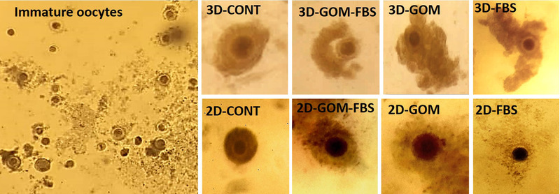
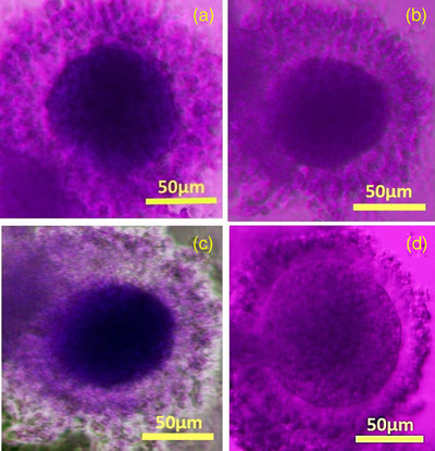
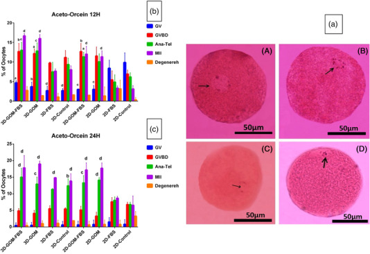
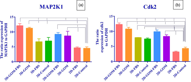
Cumulus-oocyte complexes were recovered from rabbits and divided into 3D and 2D conditions cultured for 12 and 24 h. In 3D cultures, the oocytes embedded in alginate containing FBS decellularized GOM. Corresponding supplements were also added in 2D conditions-maturation of the oocytes evaluated by Aceto-Orcein, TEM, and RT-PCR for MAP2K1 and Cdk2.
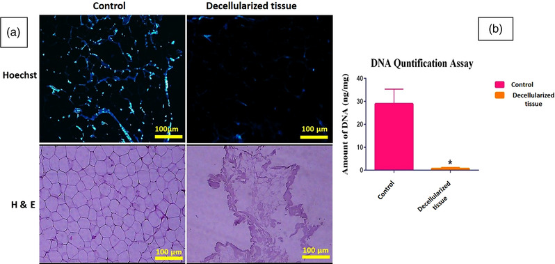
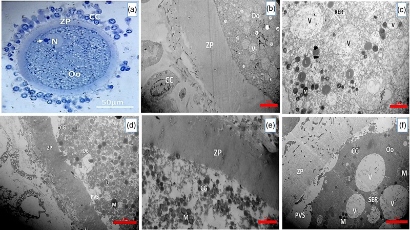
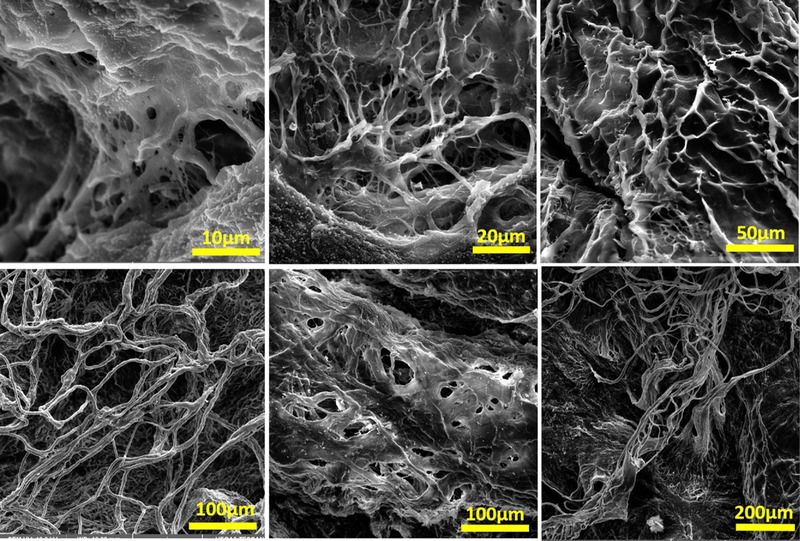
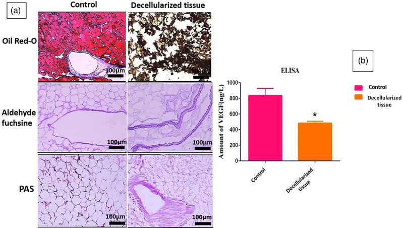
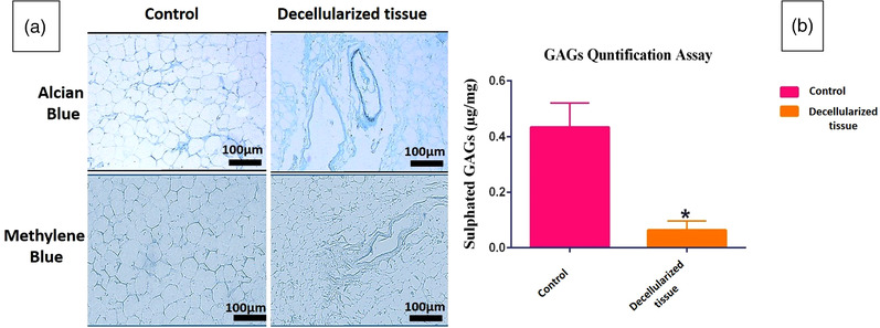
DNA quantification, Hoechst, and H&E staining confirmed cell depletion from GOM, and SEM showed the preservation of ultra-architecture after decellularization. Histochemical staining methods showed appropriate extracellular matrix preservation. ELISA assessment showed retention of VEGF content. MTT assessment indicated decellularized GOM was non-toxic. Both Aceto-Orcein assessment and ultra-structure study of the oocytes showed that supplementation of 2D or 3D cultures with decellularized omentum promoted oocyte maturation. Expression of MAP2K1 and Cdk2 also increased in the presence of GOM.
GOM supplementation has a beneficial impact on oocyte maturation, probably due to the presence of growth factors and proteins.
为了预防卵巢过度刺激综合征,体外成熟(IVM)允许卵母细胞在没有激素治疗的情况下进行不孕治疗。虽然在 IVM 过程中成熟了许多卵母细胞,但一些培养条件的缺陷导致卵丘细胞和卵母细胞核和细胞质成熟的生长和发育受到抑制。
改善卵母细胞培养条件的挑战促使我们使用富含生长因子和蛋白质的大网膜(GOM)作为基础培养培养基的丰富补充。
从兔子中回收卵丘-卵母细胞复合物,并分为 3D 和 2D 条件培养 12 和 24 小时。在 3D 培养中,将卵母细胞嵌入含有 FBS 的藻酸盐中,其中包含去细胞化的 GOM。在 2D 条件下也添加了相应的补充物——通过 Aceto-Orcein、TEM 和 RT-PCR 评估卵母细胞的成熟情况,以评估 MAP2K1 和 Cdk2。
DNA 定量、Hoechst 和 H&E 染色证实了 GOM 中的细胞耗竭,SEM 显示去细胞化后超微结构得以保留。组织化学染色方法显示适当的细胞外基质保留。ELISA 评估显示 VEGF 含量得以保留。MTT 评估表明去细胞化的 GOM 没有毒性。Aceto-Orcein 评估和卵母细胞的超微结构研究均表明,2D 或 3D 培养中添加去细胞化的大网膜可促进卵母细胞成熟。MAP2K1 和 Cdk2 的表达也在 GOM 的存在下增加。
GOM 的补充对卵母细胞成熟有有益的影响,这可能是由于生长因子和蛋白质的存在。