Arizona State University-Banner Neurodegenerative Disease Research Center at the Biodesign Institute, Arizona State University, 797 E Tyler St, Tempe, AZ, 85287, USA.
School of Life Sciences, Arizona State University, Tempe, AZ, USA.
J Neuroinflammation. 2022 Jul 28;19(1):193. doi: 10.1186/s12974-022-02544-5.
Herbicides are environmental contaminants that have gained much attention due to the potential hazards they pose to human health. Glyphosate, the active ingredient in many commercial herbicides, is the most heavily applied herbicide worldwide. The recent rise in glyphosate application to corn and soy crops correlates positively with increased death rates due to Alzheimer's disease and other neurodegenerative disorders. Glyphosate has been shown to cross the blood-brain barrier in in vitro models, but has yet to be verified in vivo. Additionally, reports have shown that glyphosate exposure increases pro-inflammatory cytokines in blood plasma, particularly TNFα.
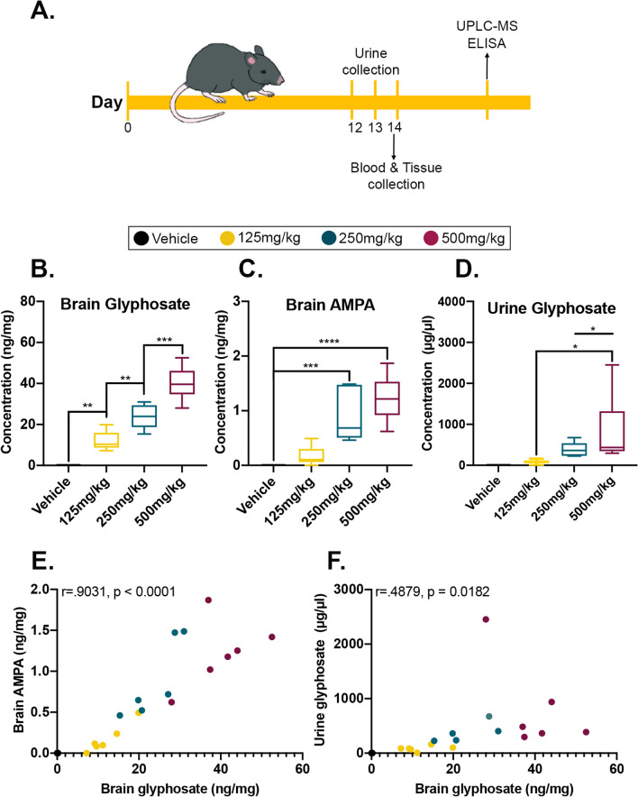
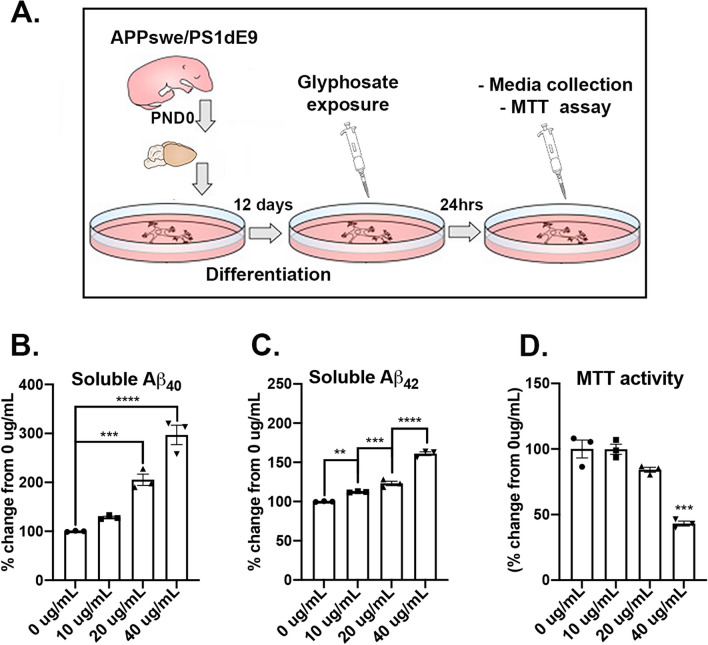
Here, we examined whether glyphosate infiltrates the brain and elevates TNFα levels in 4-month-old C57BL/6J mice. Mice received either 125, 250, or 500 mg/kg/day of glyphosate, or a vehicle via oral gavage for 14 days. Urine, plasma, and brain samples were collected on the final day of dosing for analysis via UPLC-MS and ELISAs. Primary cortical neurons were derived from amyloidogenic APP/PS1 pups to evaluate in vitro changes in Aβ burden and cytotoxicity. RNA sequencing was performed on C57BL/6J brain samples to determine changes in the transcriptome.
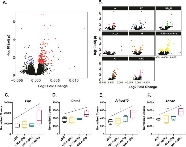
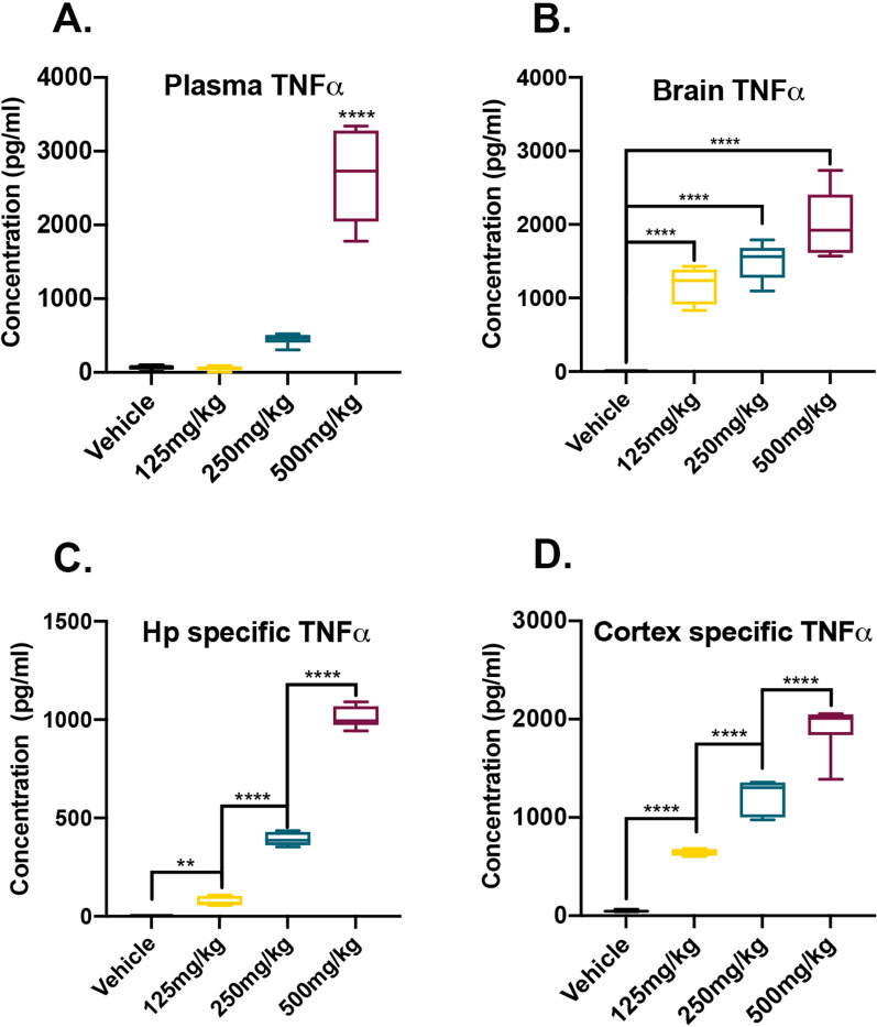
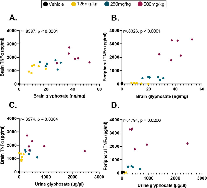
Our analysis revealed that glyphosate infiltrated the brain in a dose-dependent manner and upregulated TNFα in both plasma and brain tissue post-exposure. Notably, glyphosate measures correlated positively with TNFα levels. Glyphosate exposure in APP/PS1 primary cortical neurons increases levels of soluble Aβ and cytotoxicity. RNAseq revealed over 200 differentially expressed genes in a dose-dependent manner and cell-type-specific deconvolution analysis showed enrichment of key biological processes in oligodendrocytes including myelination, axon ensheathment, glial cell development, and oligodendrocyte development.
Collectively, these results show for the first time that glyphosate infiltrates the brain, elevates both the expression of TNFα and soluble Aβ, and disrupts the transcriptome in a dose-dependent manner, suggesting that exposure to this herbicide may have detrimental outcomes regarding the health of the general population.
除草剂是环境污染物,由于其对人类健康构成的潜在危害,引起了广泛关注。草甘膦是许多商业除草剂中的有效成分,是全球应用最广泛的除草剂。最近,玉米和大豆作物中草甘膦的使用量增加,与阿尔茨海默病和其他神经退行性疾病导致的死亡率上升呈正相关。在体外模型中,草甘膦已被证明可以穿过血脑屏障,但尚未在体内得到证实。此外,有报告显示,草甘膦暴露会增加血浆中的促炎细胞因子,尤其是 TNFα。
在这里,我们研究了草甘膦是否会渗透到大脑中,并在 4 个月大的 C57BL/6J 小鼠中升高 TNFα 水平。将小鼠通过口服灌胃接受 125、250 或 500mg/kg/天的草甘膦或载体,共 14 天。在最后一天给药时收集尿液、血浆和脑组织样本,通过 UPLC-MS 和 ELISA 进行分析。从淀粉样蛋白生成 APP/PS1 幼崽中分离出原代皮质神经元,以评估 Aβ 负荷和细胞毒性的体外变化。对 C57BL/6J 大脑样本进行 RNA 测序,以确定转录组的变化。
我们的分析表明,草甘膦以剂量依赖的方式渗透到大脑中,并在暴露后同时上调血浆和脑组织中的 TNFα。值得注意的是,草甘膦的测量值与 TNFα 水平呈正相关。APP/PS1 原代皮质神经元中的草甘膦暴露会增加可溶性 Aβ 的水平并导致细胞毒性。RNAseq 以剂量依赖的方式显示出 200 多个差异表达基因,细胞类型特异性去卷积分析显示在少突胶质细胞中丰富的关键生物学过程,包括髓鞘形成、轴突包裹、神经胶质细胞发育和少突胶质细胞发育。
总的来说,这些结果首次表明,草甘膦渗透到大脑中,以剂量依赖的方式升高 TNFα 和可溶性 Aβ 的表达,并破坏转录组,表明接触这种除草剂可能对一般人群的健康产生不利影响。