Viswan Parvathy, Behera Biswanath, Sethy Madhusmita, Dash Siddhartha, Palit Aparna, Ayyanar Pavithra
Dermatology, Venereology and Leprology, All India Institute of Medical Sciences, Bhubaneswar, Bhubaneswar, IND.
Pathology and Laboratory Medicine, All India Institute of Medical Sciences, Bhubaneswar, Bhubaneswar, IND.
Cureus. 2022 Jun 24;14(6):e26292. doi: 10.7759/cureus.26292. eCollection 2022 Jun.
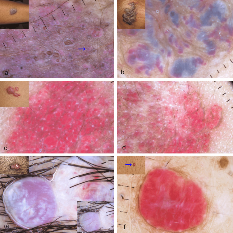
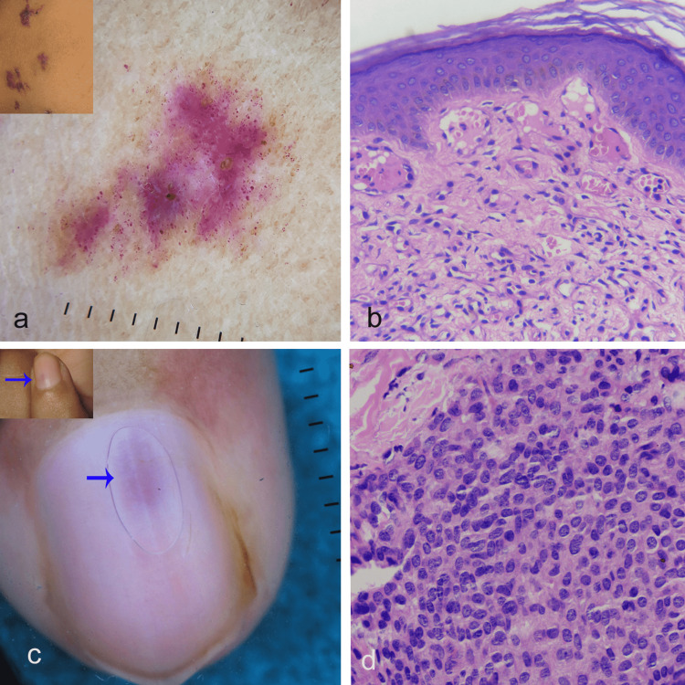
Background Cutaneous vascular malformations and tumors comprise a vast group of conditions with variable clinical presentations. It is imperative to differentiate them from nonvascular lesions and from each other as their management and prognosis differ significantly. There is only sparse literature on dermoscopic features of various vascular malformations and tumors, especially from India. Aim We aimed to retrospectively study the dermoscopic findings of various vascular malformations and tumors based on their dominant vascular dermoscopic feature. Method All the vascular malformations and tumors for which clinical details and clinical and dermoscopic images were available were included in the analysis. The dominant vascular feature(s) was defined as a single or combination of two or more vascular features (in case a single vascular feature does not satisfy the criteria) that constitute more than 75% of the lesions' vascular features. These included red, purple, blue, black (or any combination) dots, globules, lacunae, structureless area, linear, linear irregular, hairpin, comma, and arborizing vessels. Results A total of 52 patients with 68 vascular lesions (22 vascular malformations and 46 vascular tumors) were analyzed. Port-wine stain showed linear irregular vessels with sharp border with or without intervening white structureless area; unilateral nevoid telangiectasia had red dots and globules; angiokeratoma displayed red, reddish-purple to brown lacunae; blue color was seen in venous and glomuvenous malformation and venous lake; a mixed pattern was noted in infantile hemangioma and verrucous hemangioma; a red to reddish-white structureless area was observed in pyogenic granuloma and cherry angioma, and a subungual ill-defined pink structureless area was spotted in subungual glomus tumor. Conclusion The dermoscopic features observed in various vascular lesions may overlap; however, the dominant dermoscopic feature along with its color may point to the diagnosis.
皮肤血管畸形和肿瘤包含一大类临床表现各异的病症。鉴于其治疗方法和预后差异显著,将它们与非血管性病变以及彼此区分开来至关重要。关于各种血管畸形和肿瘤的皮肤镜特征的文献稀少,尤其是来自印度的。
我们旨在基于各种血管畸形和肿瘤的主要血管皮肤镜特征进行回顾性研究。
分析所有具备临床细节以及临床和皮肤镜图像的血管畸形和肿瘤。主要血管特征被定义为构成病变血管特征75%以上的单个血管特征或两个或更多血管特征的组合(如果单个血管特征不符合标准)。这些特征包括红色、紫色、蓝色、黑色(或任何组合)的点状、球状、腔隙、无结构区域、线状、线状不规则、发夹状、逗号状和树枝状血管。
共分析了52例患者的68处血管病变(22处血管畸形和46处血管肿瘤)。葡萄酒色斑显示线状不规则血管,边界清晰,有或无中间白色无结构区域;单侧痣样毛细血管扩张有红色点状和球状;血管角化瘤显示红色、红紫色至棕色腔隙;静脉畸形、球静脉畸形和静脉湖可见蓝色;婴儿血管瘤和疣状血管瘤可见混合模式;化脓性肉芽肿和樱桃状血管瘤观察到红色至红白色无结构区域,甲下球瘤可见甲下边界不清的粉红色无结构区域。
在各种血管病变中观察到的皮肤镜特征可能会重叠;然而,主要的皮肤镜特征及其颜色可能有助于诊断。