Touarsa Firdaous, El Ouali Ibtissam, Elkettani Najwa, Fikri Meriem, Jiddane Mohamed
Neuroradiology Department, Specialty Hospital of Rabat, Rabat, Morocco.
SAGE Open Med Case Rep. 2022 Jul 22;10:2050313X221113261. doi: 10.1177/2050313X221113261. eCollection 2022.
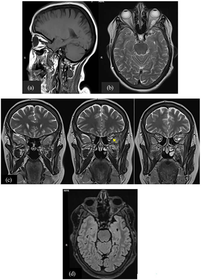
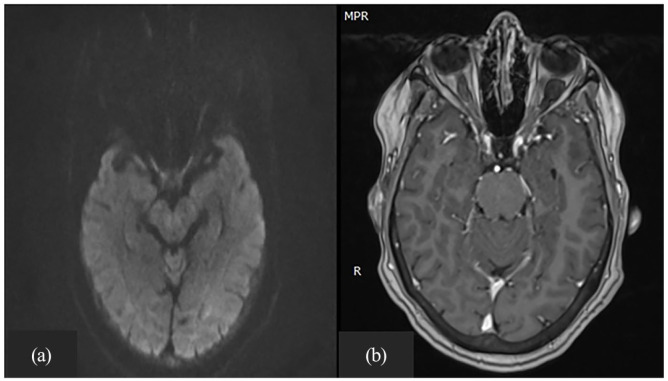
Arachnoid cysts are the most common benign cystic abnormalities formed due to congenital splitting of the arachnoid layer. They comprise 1% of intracranial masses, and the orbital location is even more rarely reported in history especially in the pediatric population. They might be discovered as an asymptomatic finding on imaging performed for a concomitant condition or, in most reported cases, as a result of ophthalmic impairment. They can be isolated or associated with gliomas, neurofibromas, empty sella syndrome, and frontotemporal porencephalic cysts. Computed tomography scan shows a non-enhancing liquid cystic lesion, and magnetic resonance imaging remains the best assessment tool confirming the similarity of the fluid to cerebrospinal fluid and evaluating the optic nerves. Herein, we report the case of an incidental discovery of an intraorbital arachnoid cyst on magnetic resonance imaging in a 53-year-old woman with a history of epilepsy. No treatment was performed as the cystic formation was asymptomatic.
蛛网膜囊肿是由于蛛网膜层先天性分裂形成的最常见的良性囊性异常。它们占颅内肿块的1%,而眼眶部位在历史上报道更为罕见,尤其是在儿科人群中。它们可能在因伴随疾病进行的影像学检查中作为无症状发现被发现,或者在大多数报道的病例中,是由于眼部损伤而被发现。它们可以是孤立的,也可与胶质瘤、神经纤维瘤、空蝶鞍综合征和额颞叶脑穿通畸形囊肿相关。计算机断层扫描显示为无强化的液性囊性病变,而磁共振成像仍然是确认液体与脑脊液相似性并评估视神经的最佳评估工具。在此,我们报告一例53岁有癫痫病史的女性在磁共振成像时偶然发现眶内蛛网膜囊肿的病例。由于囊性结构无症状,未进行治疗。