Pathology and Forensic Medicine Department, Federal University of Ceará, Fortaleza, CE, Brazil.
Laboratory of Genomic Studies, Biology Department, São Paulo State University (UNESP), São José do Rio Preto, SP, Brazil.
Asian Pac J Cancer Prev. 2022 Jul 1;23(7):2351-2359. doi: 10.31557/APJCP.2022.23.7.2351.
This study aimed to determine the presence of Epstein-Barr Virus (EBV) and Human papillomavirus (HPV) in breast cancer with patients from Northeast of Brazil, considering the molecular subtypes and also taking in account the relation with TP53 immunoexpression.
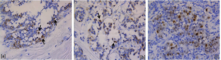
Seventy-five samples of invasive breast carcinoma with no special type were selected from pathology archives at Federal University of Ceará. EBV was detected by In situ hybridization (ISH) and immunohistochemistry (IHC) and HPV was detected by PCR. ISH was performed using EBER1 probe (Shibata et al., 1991; Bacchi et al., 1996) while IHC was performed on histological formalin-fixed paraffin-embedded tissue samples (Hsu et al., 1981). PCR methodology (Haws et al., 2004) was used to amplify the genetic material of human papillomavirus. The amplification products were electrophoretic analyzed on 1% agarose gel. The data analyses were carried out using the statistical software EPINFO® version 6.04d and SPSS version 17.0 (SPSS Inc., Chicago, IL). Statistically significant differences were evaluated by the chi-square test and Fisher's exact test and correlations between groups were analyzed by Spearman's and Pearson's rank correlation coefficient.
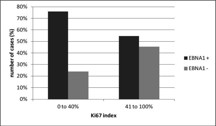
69.4% of the cases were EBNA1 positives by IHC. EBNA1 positive tumors had lower Ki-67 index (0-40%), while EBNA1 negative cases had relevant higher Ki-67 index (41-100%) (p = 0.06). EBV was present in all tumor grades, with a high frequency in grade I and III tumors comparing to EBNA1 negative cases. No HPV positive cases were observed.
Regarding the results from this study, we support the hypothesis that EBV can be involved on breast tumorigenesis.
本研究旨在确定巴西东北部乳腺癌患者中 Epstein-Barr 病毒(EBV)和人乳头瘤病毒(HPV)的存在情况,同时考虑到分子亚型,并考虑与 TP53 免疫表达的关系。
从联邦大学塞阿拉病理学档案中选择了 75 例无特殊类型的浸润性乳腺癌病例。通过原位杂交(ISH)和免疫组织化学(IHC)检测 EBV,通过聚合酶链反应(PCR)检测 HPV。ISH 使用 EBER1 探针(Shibata 等人,1991 年;Bacchi 等人,1996 年)进行,而 IHC 则在组织学福尔马林固定石蜡包埋组织样本(Hsu 等人,1981 年)上进行。使用聚合酶链反应方法(Haws 等人,2004 年)扩增人乳头瘤病毒的遗传物质。扩增产物在 1%琼脂糖凝胶上进行电泳分析。使用统计软件 EPINFO®版本 6.04d 和 SPSS 版本 17.0(SPSS Inc.,芝加哥,IL)进行数据分析。使用卡方检验和 Fisher 确切检验进行统计学显著性差异评估,使用 Spearman 和 Pearson 等级相关系数分析组间相关性。
69.4%的病例通过 IHC 检测到 EBNA1 阳性。EBNA1 阳性肿瘤的 Ki-67 指数(0-40%)较低,而 EBNA1 阴性病例的 Ki-67 指数(41-100%)较高(p=0.06)。EBV 存在于所有肿瘤分级中,与 EBNA1 阴性病例相比,I 级和 III 级肿瘤的 EBV 存在频率更高。未观察到 HPV 阳性病例。
根据本研究结果,我们支持 EBV 可能参与乳腺癌发生的假说。