• 文献检索
  • 文档翻译
  • 深度研究
  • 学术资讯
  • Suppr Zotero 插件Zotero 插件
  • 邀请有礼
  • 套餐&价格
  • 历史记录
应用&插件
Suppr Zotero 插件Zotero 插件浏览器插件Mac 客户端Windows 客户端微信小程序
定价
高级版会员购买积分包购买API积分包
服务
文献检索文档翻译深度研究API 文档MCP 服务
关于我们
关于 Suppr公司介绍联系我们用户协议隐私条款
关注我们

Suppr 超能文献

核心技术专利:CN118964589B侵权必究
粤ICP备2023148730 号-1Suppr @ 2026

文献检索

告别复杂PubMed语法,用中文像聊天一样搜索,搜遍4000万医学文献。AI智能推荐,让科研检索更轻松。

立即免费搜索

文件翻译

保留排版,准确专业,支持PDF/Word/PPT等文件格式,支持 12+语言互译。

免费翻译文档

深度研究

AI帮你快速写综述,25分钟生成高质量综述,智能提取关键信息,辅助科研写作。

立即免费体验

视网膜病变眼中的视网膜外层褶皱的临床和影像学特征。

Clinical and imaging characteristics of outer retinal folds in eyes with retinitis.

机构信息

Uveitis and Retina Services, L V Prasad Eye Institute, Mithu Tulsi Chanrai Campus, Bhubaneswar, Odisha, India.

Uveitis and Retina Services, L V Prasad Eye Institute, Mithu Tulsi Chanrai Campus, Bhubaneswar, Odisha; Uveitis and Retina Services, C L Gupta Eye Institute, Moradabad, Uttar Pradesh, India.

出版信息

Indian J Ophthalmol. 2022 Aug;70(8):2981-2985. doi: 10.4103/ijo.IJO_70_22.

DOI:10.4103/ijo.IJO_70_22
PMID:35918957
原文链接:https://pmc.ncbi.nlm.nih.gov/articles/PMC9672790/
Abstract

PURPOSE

To describe clinical and imaging characteristics of the outer retinal folds (ORF) in cases of retinitis, retinochoroiditis, and chorioretinitis.

METHODS

Retrospective review of retinitis cases with presence of ORFs either at presentation or during follow up.

RESULTS

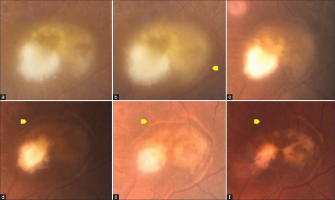
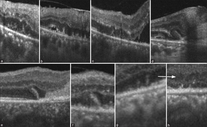
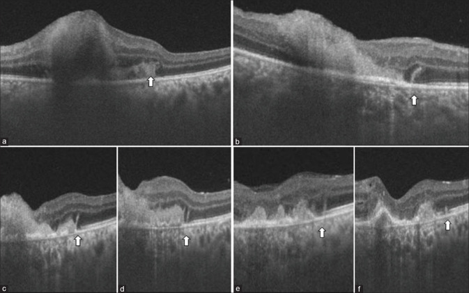
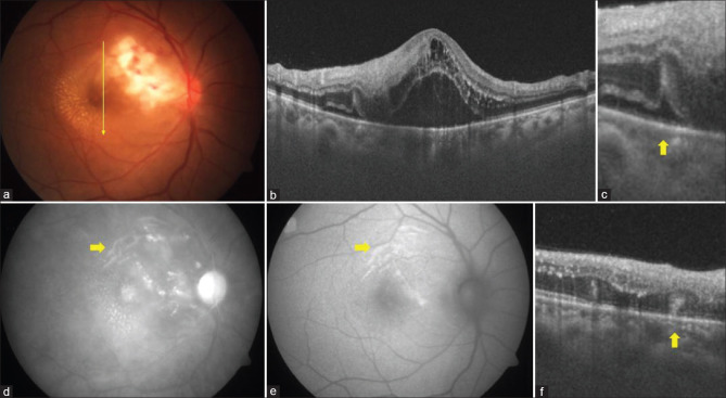
ORFs were seen adjacent to retinitis lesions in 16 eyes of 14 cases (retinitis post-febrile illness n = 10, toxoplasma retinochoroiditis n = 2, fungal chorioretinitis n = 2) either at presentation (n = 2) or during follow up (n = 14). Optical coherence tomography (OCT) appearance was outer retinal vertical stout lesions involving ellipsoid, external limiting membrane, and outer nuclear layer. All the cases had a presence of past or concurrent subretinal fluid and/or subretinal hyperreflective material when ORF was seen. ORF resolved with variable outer retinal atrophy over a mean period of 2.86 months.

CONCLUSION

ORF is observed in cases of retinitis with subretinal fluid either at presentation or during resolution. It is not specific to any etiological disease. Differentiation of this sign from vertical outer retinal stripes in viral retinitis on OCT is important to avoid misinterpretation.

摘要

目的

描述视网膜病变、脉络膜炎和脉络膜视网膜炎病例中外层视网膜褶皱(ORF)的临床和影像学特征。

方法

回顾性分析存在 ORF 的视网膜病变病例,这些 ORF 或在就诊时出现,或在随访期间出现。

结果

在 14 例病例的 16 只眼中(发热后性视网膜病变 n = 10,弓形体脉络膜视网膜炎 n = 2,真菌性脉络膜炎 n = 2),无论是在就诊时(n = 2)还是在随访期间(n = 14),均发现 ORF 紧邻视网膜病变病灶。光学相干断层扫描(OCT)表现为累及椭圆体、外界膜和外核层的垂直粗壮的外层视网膜病变。当出现 ORF 时,所有病例均存在既往或同时存在的视网膜下液和/或视网膜下高反射物质。ORF 在平均 2.86 个月的时间内,随着可变的外层视网膜萎缩而消退。

结论

在外层视网膜下液存在于就诊时或消退期间的视网膜病变病例中观察到 ORF。它并非特定于任何病因疾病。在 OCT 上,将此征象与病毒性视网膜病变中的垂直外视网膜条纹区分开来非常重要,以避免误判。

https://cdn.ncbi.nlm.nih.gov/pmc/blobs/69ae/9672790/c6855b67dd24/IJO-70-2981-g004.jpg
https://cdn.ncbi.nlm.nih.gov/pmc/blobs/69ae/9672790/39cb1f13dd42/IJO-70-2981-g001.jpg
https://cdn.ncbi.nlm.nih.gov/pmc/blobs/69ae/9672790/d232cc4ff3c4/IJO-70-2981-g002.jpg
https://cdn.ncbi.nlm.nih.gov/pmc/blobs/69ae/9672790/f8a2cc676caf/IJO-70-2981-g003.jpg
https://cdn.ncbi.nlm.nih.gov/pmc/blobs/69ae/9672790/c6855b67dd24/IJO-70-2981-g004.jpg

相似文献

1
Clinical and imaging characteristics of outer retinal folds in eyes with retinitis.视网膜病变眼中的视网膜外层褶皱的临床和影像学特征。
Indian J Ophthalmol. 2022 Aug;70(8):2981-2985. doi: 10.4103/ijo.IJO_70_22.
2
Spectral domain optical coherence tomography findings in patients with acute syphilitic posterior placoid chorioretinopathy.急性梅毒性后浆性脉络膜视网膜炎患者的光谱域光学相干断层扫描结果。
Retina. 2014 Feb;34(2):373-84. doi: 10.1097/IAE.0b013e3182993f11.
3
SWEPT-SOURCE OPTICAL COHERENCE TOMOGRAPHY ANGIOGRAPHY IN RICKETTSIAL RETINITIS.立克次体性视网膜炎的扫频源光学相干断层扫描血管造影术
Retin Cases Brief Rep. 2019 Fall;13(4):348-351. doi: 10.1097/ICB.0000000000000603.
4
Multimodal imaging in a case of bilateral outer retinitis associated with mumps infection.一例与腮腺炎感染相关的双侧外层视网膜炎的多模态成像
Int Ophthalmol. 2018 Feb;38(1):339-343. doi: 10.1007/s10792-016-0417-y. Epub 2016 Dec 27.
5
EN FACE OPTICAL COHERENCE TOMOGRAPHY AND OPTICAL COHERENCE TOMOGRAPHY ANGIOGRAPHY OF MULTIPLE EVANESCENT WHITE DOT SYNDROME: New Insights Into Pathogenesis.多灶性易消散性白点综合征的正面光学相干断层扫描和光学相干断层扫描血管造影:发病机制的新见解
Retina. 2016 Dec;36 Suppl 1:S178-S188. doi: 10.1097/IAE.0000000000001255.
6
OPTICAL COHERENCE TOMOGRAPHY FINDINGS IN CYTOMEGALOVIRUS RETINITIS: A Longitudinal Study.巨细胞病毒性视网膜炎的光学相干断层扫描结果:一项纵向研究。
Retina. 2018 Jan;38(1):108-117. doi: 10.1097/IAE.0000000000001503.
7
Acute retinal pigment epitheliitis: spectral-domain optical coherence tomography findings in 18 cases.急性视网膜色素上皮炎:18 例的频域光相干断层扫描表现。
Invest Ophthalmol Vis Sci. 2014 May 1;55(5):3314-9. doi: 10.1167/iovs.14-14324.
8
Optical Coherence Tomography Findings in Endogenous Fungal Chorioretinitis, Retinitis, and Endophthalmitis.内源性真菌性脉络膜视网膜炎、视网膜炎和眼内炎的光学相干断层扫描结果
Ophthalmic Surg Lasers Imaging Retina. 2017 Nov 1;48(11):894-901. doi: 10.3928/23258160-20171030-04.
9
MEASUREMENTS OF RETINAL FLUID BY OPTICAL COHERENCE TOMOGRAPHY LEAKAGE IN DIABETIC MACULAR EDEMA: A Biomarker of Visual Acuity Response to Treatment.光学相干断层扫描渗漏测量在糖尿病性黄斑水肿中的应用:视力反应治疗的生物标志物。
Retina. 2019 Jan;39(1):52-60. doi: 10.1097/IAE.0000000000001905.
10
Multimodal imaging in persistent placoid maculopathy.持久性盘状斑状病变的多模态影像学。
JAMA Ophthalmol. 2014 Jan;132(1):38-49. doi: 10.1001/jamaophthalmol.2013.6310.

引用本文的文献

1
Post-fever Retinitis With a Positive Weil-Felix Test: A Study From a Tertiary Center in South India.发热后视网膜病变伴外斐试验阳性:来自印度南部一家三级中心的研究
Cureus. 2024 Jan 29;16(1):e53162. doi: 10.7759/cureus.53162. eCollection 2024 Jan.

本文引用的文献

1
Outer Retinal Folds after Pars Plana Vitrectomy vs. Pneumatic Retinopexy for Retinal Detachment Repair: Post hoc analysis from PIVOT.视网膜脱离修复中玻璃体切除联合视网膜光凝术与气体视网膜固定术术后视网膜外层褶皱:PIVOT研究的事后分析
Ophthalmol Retina. 2022 Mar;6(3):234-242. doi: 10.1016/j.oret.2021.09.001. Epub 2021 Sep 11.
2
Optical coherence tomography diagnostic signs in posterior uveitis.后葡萄膜炎的光学相干断层扫描诊断征象。
Prog Retin Eye Res. 2020 Mar;75:100797. doi: 10.1016/j.preteyeres.2019.100797. Epub 2019 Sep 9.
3
Optical coherence tomography and optical coherence tomography angiography in uveitis: A review.
《葡萄膜炎的光学相干断层扫描和光学相干断层扫描血管成像:综述》。
Clin Exp Ophthalmol. 2019 Apr;47(3):357-371. doi: 10.1111/ceo.13470. Epub 2019 Mar 3.
4
Inner, outer, and full-thickness retinal folds after rhegmatogenous retinal detachment repair: A review.裂孔源性视网膜脱离修复术后的视网膜内、外和全层皱褶:综述。
Surv Ophthalmol. 2019 Mar-Apr;64(2):135-161. doi: 10.1016/j.survophthal.2018.10.007. Epub 2018 Nov 1.
5
Comparing optical coherence tomography findings in different aetiologies of infectious necrotising retinitis.比较不同病因感染性坏死性视网膜炎的光学相干断层扫描结果。
Br J Ophthalmol. 2018 Apr;102(4):433-437. doi: 10.1136/bjophthalmol-2017-310210. Epub 2017 Aug 1.
6
The application of optical coherence tomography angiography in uveitis and inflammatory eye diseases.光学相干断层扫描血管造影在葡萄膜炎和炎症性眼病中的应用。
Prog Retin Eye Res. 2017 Jul;59:178-201. doi: 10.1016/j.preteyeres.2017.04.005. Epub 2017 Apr 29.
7
Optical coherence tomography evaluation of patients with macula-off retinal detachment after different postoperative posturing: a randomized pilot study.不同术后体位黄斑脱离视网膜脱离患者的光学相干断层扫描评估:一项随机试验研究
Acta Ophthalmol. 2017 Aug;95(5):e379-e384. doi: 10.1111/aos.13397. Epub 2017 Apr 26.
8
Evolution of outer retinal folds occurring after vitrectomy for retinal detachment repair.视网膜脱离修复玻璃体切除术后外视网膜皱襞的演变。
Invest Ophthalmol Vis Sci. 2012 Dec 3;53(13):7928-35. doi: 10.1167/iovs.12-10322.
9
Longitudinal study of macular folds by spectral-domain optical coherence tomography.通过光谱域光学相干断层扫描对黄斑褶皱进行的纵向研究。
Am J Ophthalmol. 2012 Oct;154(4):757-8; author reply 758-9. doi: 10.1016/j.ajo.2012.06.013.
10
Longitudinal study of macular folds by spectral-domain optical coherence tomography.频域光学相干断层扫描对黄斑皱褶的纵向研究。
Am J Ophthalmol. 2012 Jan;153(1):88-92.e1. doi: 10.1016/j.ajo.2011.06.003. Epub 2011 Aug 19.