• 文献检索
  • 文档翻译
  • 深度研究
  • 学术资讯
  • Suppr Zotero 插件Zotero 插件
  • 邀请有礼
  • 套餐&价格
  • 历史记录
应用&插件
Suppr Zotero 插件Zotero 插件浏览器插件Mac 客户端Windows 客户端微信小程序
定价
高级版会员购买积分包购买API积分包
服务
文献检索文档翻译深度研究API 文档MCP 服务
关于我们
关于 Suppr公司介绍联系我们用户协议隐私条款
关注我们

Suppr 超能文献

核心技术专利:CN118964589B侵权必究
粤ICP备2023148730 号-1Suppr @ 2026

文献检索

告别复杂PubMed语法,用中文像聊天一样搜索,搜遍4000万医学文献。AI智能推荐,让科研检索更轻松。

立即免费搜索

文件翻译

保留排版,准确专业,支持PDF/Word/PPT等文件格式,支持 12+语言互译。

免费翻译文档

深度研究

AI帮你快速写综述,25分钟生成高质量综述,智能提取关键信息,辅助科研写作。

立即免费体验

与高分辨率计算机断层扫描相比,三维超短回波时间磁共振成像在特发性肺纤维化评估中的应用

Three-dimensional ultrashort echo time magnetic resonance imaging in assessment of idiopathic pulmonary fibrosis, in comparison with high-resolution computed tomography.

作者信息

Yang Xiaoyan, Liu Min, Duan Jianghui, Sun Haishuang, An Jing, Benkert Thomas, Dai Huaping, Wang Chen

机构信息

Department of Pulmonary and Critical Care Medicine, China-Japan Friendship Hospital, Capital Medical University, Beijing, China.

National Center for Respiratory Medicine, Institute of Respiratory Medicine, Chinese Academy of Medical Sciences, National Clinical Research Center for Respiratory Diseases, Beijing, China.

出版信息

Quant Imaging Med Surg. 2022 Aug;12(8):4176-4189. doi: 10.21037/qims-21-1133.

DOI:10.21037/qims-21-1133
PMID:35919053
原文链接:https://pmc.ncbi.nlm.nih.gov/articles/PMC9338383/
Abstract

BACKGROUND

We aimed to evaluate the image quality, feasibility, and diagnostic performance of three-dimensional ultrashort echo time magnetic resonance imaging (3D UTE-MRI) to assess idiopathic pulmonary fibrosis (IPF) compared with high-resolution computed tomography (HRCT) and half-Fourier single-shot turbo spin-echo (HASTE) MRI.

METHODS

A total of 36 patients with IPF (34 men; mean age: 62±8 years, age range: 43 to 78 years) were prospectively included and underwent HRCT and chest MRI on the same day. Chest MRI was performed with a free-breathing 3D spiral UTE pulse sequence and HASTE sequence on a 1.5 T MRI. Two radiologists independently evaluated the image quality of the HRCT, HASTE, and 3D UTE-MRI. They assessed the representative imaging features of IPF, including honeycombing, reticulation, traction bronchiectasis, and ground-glass opacities. Image quality of the 3D UTE-MRI, HASTE, and HRCT were assessed using a 5-point visual scoring method. Kappa and weighted kappa analysis were used to measure intra- and inter-observer and inter-method agreements. Sensitivity (SE), specificity (SP), and accuracy (AC) were used to assess the performance of 3D UTE-MRI for detecting image features of IPF and monitoring the extent of pulmonary fibrosis. Linear regressions and Bland-Altman plots were generated to assess the correlation and agreement between the assessment of the extent of pulmonary fibrosis made by the 2 observers.

RESULTS

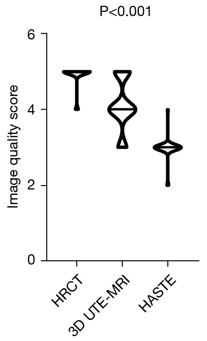
The image quality of HRCT was higher than that of HASTE and UTE-MRI (HRCT UTE-MRI HASTE: 4.9±0.3 4.1±0.7 3.0±0.3; P<0.001). Interobserver agreement of HRCT, HASTE, and 3D UTE-MRI when assessing pulmonary fibrosis was substantial and excellent (HRCT: 0.727≤ κ ≤1, P<0.001; HASTE: 0.654≤ κ ≤1, P<0.001; 3D UTE-MRI: 0.719≤ κ ≤0.824, P<0.001). In addition, reticulation (SE: 97.1%; SP: 100%; AC: 97.2%; κ =0.654), honeycombing (SE: 83.3%; SP: 100%; AC: 86.1%; κ =0.625) patterns, and traction bronchiectasis (SE: 94.1%; SP: 100%; AC: 94.4%, κ =0.640) were also well-visualized on 3D UTE-MRI, which was significantly superior to HASTE. Compared with HRCT, the sensitivity of 3D UTE-MRI to detect signs of pulmonary fibrosis (n=35) was 97.2%. The interobserver agreement in elevation of the extent of pulmonary fibrosis with HRCT and 3D UTE-MRI was R=0.84 (P<0.001) and R=0.84 (P<0.001), respectively. The extent of pulmonary fibrosis assessed with 3D UTE-MRI [median =9, interquartile range (IQR): 6.25 to 10.00] was lower than that from HRCT (median =12, IQR: 9.25 to 13.00; U=320.00, P<0.001); however, they had a positive correlation (R=0.72, P<0.001).

CONCLUSIONS

As a radiation-free non-contrast enhanced imaging method, although the image quality of 3D UTE-MRI is inferior to that of HRCT, it has high reproducibility to identify the imaging features of IPF and evaluate the extent of pulmonary fibrosis.

摘要

背景

我们旨在评估三维超短回波时间磁共振成像(3D UTE-MRI)在评估特发性肺纤维化(IPF)方面的图像质量、可行性和诊断性能,并与高分辨率计算机断层扫描(HRCT)和半傅里叶单次激发快速自旋回波(HASTE)MRI进行比较。

方法

前瞻性纳入36例IPF患者(34例男性;平均年龄:62±8岁,年龄范围:43至78岁),并于同一天接受HRCT和胸部MRI检查。在1.5T MRI上使用自由呼吸的3D螺旋UTE脉冲序列和HASTE序列进行胸部MRI检查。两名放射科医生独立评估HRCT、HASTE和3D UTE-MRI的图像质量。他们评估了IPF的代表性影像学特征,包括蜂窝状改变、网状改变、牵拉性支气管扩张和磨玻璃影。使用5分视觉评分法评估3D UTE-MRI、HASTE和HRCT的图像质量。使用Kappa和加权Kappa分析来测量观察者内和观察者间以及方法间的一致性。使用敏感性(SE)、特异性(SP)和准确性(AC)来评估3D UTE-MRI检测IPF图像特征和监测肺纤维化程度的性能。生成线性回归和Bland-Altman图以评估两名观察者对肺纤维化程度评估之间的相关性和一致性。

结果

HRCT的图像质量高于HASTE和UTE-MRI(HRCT>UTE-MRI>HASTE:4.9±0.3>4.1±0.7>3.0±0.3;P<0.001)。HRCT、HASTE和3D UTE-MRI在评估肺纤维化时观察者间一致性良好且优秀(HRCT:0.727≤κ≤1,P<0.001;HASTE:0.654≤κ≤1,P<0.001;3D UTE-MRI:0.719≤κ≤0.824,P<0.001)。此外,网状改变(SE:97.1%;SP:100%;AC:97.2%;κ =0.654)、蜂窝状改变(SE:83.3%;SP:100%;AC:86.1%;κ =0.625)和牵拉性支气管扩张(SE:94.1%;SP:100%;AC:94.4%,κ =0.640)在3D UTE-MRI上也显示良好,明显优于HASTE。与HRCT相比,3D UTE-MRI检测肺纤维化征象(n = 35)的敏感性为97.2%。HRCT和3D UTE-MRI在肺纤维化程度升高方面的观察者间一致性分别为R = 0.84(P<0.001)和R = 0.84(P<0.001)。3D UTE-MRI评估的肺纤维化程度[中位数 = 9,四分位间距(IQR):6.25至10.00]低于HRCT(中位数 = 12,IQR:9.25至13.00;U = 320.00,P<0.001);然而,它们具有正相关性(R = 0.72,P<0.001)。

结论

作为一种无辐射的非对比增强成像方法,尽管3D UTE-MRI的图像质量不如HRCT,但它在识别IPF的影像学特征和评估肺纤维化程度方面具有较高的可重复性。

https://cdn.ncbi.nlm.nih.gov/pmc/blobs/fc7c/9338383/2c483c5c42ca/qims-12-08-4176-f7.jpg
https://cdn.ncbi.nlm.nih.gov/pmc/blobs/fc7c/9338383/af10f635dcc0/qims-12-08-4176-f1.jpg
https://cdn.ncbi.nlm.nih.gov/pmc/blobs/fc7c/9338383/44162c2809da/qims-12-08-4176-f2.jpg
https://cdn.ncbi.nlm.nih.gov/pmc/blobs/fc7c/9338383/02ff1c3859dc/qims-12-08-4176-f3.jpg
https://cdn.ncbi.nlm.nih.gov/pmc/blobs/fc7c/9338383/d057c0be612f/qims-12-08-4176-f4.jpg
https://cdn.ncbi.nlm.nih.gov/pmc/blobs/fc7c/9338383/792a37c54c46/qims-12-08-4176-f5.jpg
https://cdn.ncbi.nlm.nih.gov/pmc/blobs/fc7c/9338383/f6491d168837/qims-12-08-4176-f6.jpg
https://cdn.ncbi.nlm.nih.gov/pmc/blobs/fc7c/9338383/2c483c5c42ca/qims-12-08-4176-f7.jpg

相似文献

1
Three-dimensional ultrashort echo time magnetic resonance imaging in assessment of idiopathic pulmonary fibrosis, in comparison with high-resolution computed tomography.与高分辨率计算机断层扫描相比,三维超短回波时间磁共振成像在特发性肺纤维化评估中的应用
Quant Imaging Med Surg. 2022 Aug;12(8):4176-4189. doi: 10.21037/qims-21-1133.
2
Pulmonary Imaging of Immunocompromised Patients during Hematopoietic Stem Cell Transplantation using Non-Contrast-Enhanced Three-Dimensional Ultrashort Echo Time (3D-UTE) MRI.使用非对比增强三维超短回波时间(3D-UTE)磁共振成像对造血干细胞移植期间免疫功能低下患者进行肺部成像
Rofo. 2022 Jan;194(1):39-48. doi: 10.1055/a-1535-2341. Epub 2021 Oct 14.
3
Detection of solid and subsolid pulmonary nodules with lung MRI: performance of UTE, T1 gradient-echo, and single-shot T2 fast spin echo.肺 MRI 检测实性和亚实性肺结节:UTE、T1 梯度回波和单次激发 T2 快速自旋回波的性能。
Cancer Imaging. 2023 Feb 16;23(1):17. doi: 10.1186/s40644-023-00531-4.
4
Assessment of lung deformation in patients with idiopathic pulmonary fibrosis with elastic registration technique on pulmonary three-dimensional ultrashort echo time MRI.利用肺部三维超短回波时间磁共振成像的弹性配准技术评估特发性肺纤维化患者的肺变形情况。
Insights Imaging. 2024 Jan 23;15(1):17. doi: 10.1186/s13244-023-01555-x.
5
Assessment of chest high-field magnetic resonance imaging in children and young adults with noncystic fibrosis chronic lung disease: comparison to high-resolution computed tomography and correlation with pulmonary function.非囊性纤维化慢性肺病儿童和青年的胸部高场磁共振成像评估:与高分辨率计算机断层扫描的比较及与肺功能的相关性
Invest Radiol. 2009 Sep;44(9):532-8. doi: 10.1097/RLI.0b013e3181b4c1ba.
6
Comparison of diagnostic quality of 3D ultrashort-echo-time techniques for pulmonary magnetic resonance imaging in free-breathing.在自由呼吸状态下,比较 3D 超短回波时间技术在肺部磁共振成像中的诊断质量。
Acta Radiol. 2023 May;64(5):1851-1858. doi: 10.1177/02841851231151366. Epub 2023 Jan 30.
7
Feasibility of chest spiral 3D ultrashort echo time magnetic resonance imaging for intrathoracic metastasis work-up in breast cancer.胸部螺旋3D超短回波时间磁共振成像用于乳腺癌胸内转移评估的可行性
J Thorac Dis. 2023 Oct 31;15(10):5485-5493. doi: 10.21037/jtd-23-1006. Epub 2023 Sep 25.
8
Free-breathing ultrashort echo time lung magnetic resonance imaging using stack-of-spirals acquisition: A feasibility study in oncology patients.采用螺旋堆叠采集的自由呼吸超短回波时间肺部磁共振成像:肿瘤患者的可行性研究
Magn Reson Imaging. 2018 Sep;51:137-143. doi: 10.1016/j.mri.2018.05.002. Epub 2018 May 15.
9
Clinical Potential of UTE-MRI for Assessing COVID-19: Patient- and Lesion-Based Comparative Analysis.UTE-MRI 在评估 COVID-19 方面的临床潜力:基于患者和病变的对比分析。
J Magn Reson Imaging. 2020 Aug;52(2):397-406. doi: 10.1002/jmri.27208. Epub 2020 Jun 3.
10
Accelerated Stack-of-Spirals Free-Breathing Three-Dimensional Ultrashort Echo Time Lung Magnetic Resonance Imaging: A Feasibility Study in Patients With Breast Cancer.加速螺旋堆叠式自由呼吸三维超短回波时间肺部磁共振成像:一项针对乳腺癌患者的可行性研究
Front Oncol. 2021 Oct 7;11:746059. doi: 10.3389/fonc.2021.746059. eCollection 2021.

引用本文的文献

1
Evaluation of respiratory muscle dysfunction in patients with idiopathic pulmonary fibrosis: a prospective observational study with magnetic resonance imaging.特发性肺纤维化患者呼吸肌功能障碍的评估:一项磁共振成像的前瞻性观察研究。
BMC Pulm Med. 2025 Mar 14;25(1):118. doi: 10.1186/s12890-025-03572-6.
2
Inversion-recovery ultrashort-echo-time (IR-UTE) MRI-based detection of radiation dose heterogeneity in gynecologic cancer patients treated with HDR brachytherapy.基于反转恢复超短回波时间(IR-UTE)MRI 的宫颈癌调强近距离后装治疗中不同剂量区的检测。
Radiat Oncol. 2024 Aug 6;19(1):105. doi: 10.1186/s13014-024-02499-2.
3

本文引用的文献

1
Hyperpolarized Xe MRI and Spectroscopy of Gas-Exchange Abnormalities in Nonspecific Interstitial Pneumonia.极化氙磁共振成像与非特异性间质性肺炎气体交换异常的光谱研究。
Radiology. 2021 Oct;301(1):211-220. doi: 10.1148/radiol.2021204149. Epub 2021 Jul 27.
2
Clinical application of ultrashort echo-time MRI for lung pathologies in children.儿童肺部病变的超短回波时间 MRI 的临床应用。
Clin Radiol. 2021 Sep;76(9):708.e9-708.e17. doi: 10.1016/j.crad.2021.05.015. Epub 2021 Jun 10.
3
Chest MRI to diagnose early diaphragmatic weakness in Pompe disease.
Assessment of lung deformation in patients with idiopathic pulmonary fibrosis with elastic registration technique on pulmonary three-dimensional ultrashort echo time MRI.
利用肺部三维超短回波时间磁共振成像的弹性配准技术评估特发性肺纤维化患者的肺变形情况。
Insights Imaging. 2024 Jan 23;15(1):17. doi: 10.1186/s13244-023-01555-x.
4
Elastic Registration Algorithm Based on Three-dimensional Pulmonary MRI in Quantitative Assessment of Severity of Idiopathic Pulmonary Fibrosis.基于三维肺部磁共振成像的弹性配准算法在特发性肺纤维化严重程度定量评估中的应用
J Thorac Imaging. 2023 Sep 6;38(6):358-66. doi: 10.1097/RTI.0000000000000735.
胸部 MRI 诊断庞贝病早期膈肌无力。
Orphanet J Rare Dis. 2021 Jan 7;16(1):21. doi: 10.1186/s13023-020-01627-x.
4
Quantification of MRI T2 Interstitial Lung Disease Signal-Intensity Volume in Idiopathic Pulmonary Fibrosis: A Pilot Study.特发性肺纤维化 MRI T2 间质肺病信号强度容积的定量分析:一项初步研究。
J Magn Reson Imaging. 2021 May;53(5):1500-1507. doi: 10.1002/jmri.27454. Epub 2020 Nov 25.
5
Lung magnetic resonance imaging in systemic sclerosis: a new promising approach to evaluate pulmonary involvement and progression.系统性硬皮病的肺部磁共振成像:评估肺部受累和进展的新有前途方法。
Clin Rheumatol. 2021 May;40(5):1903-1912. doi: 10.1007/s10067-020-05491-9. Epub 2020 Nov 7.
6
Three-dimensional Ultrashort Echo Time MRI for Functional Lung Imaging in Cystic Fibrosis.三维超短回波时间 MRI 在囊性纤维化中的功能性肺部成像。
Radiology. 2020 Jul;296(1):191-199. doi: 10.1148/radiol.2020192251. Epub 2020 Apr 28.
7
Extreme MRI: Large-scale volumetric dynamic imaging from continuous non-gated acquisitions.极限 MRI:连续非门控采集的大规模容积动态成像。
Magn Reson Med. 2020 Oct;84(4):1763-1780. doi: 10.1002/mrm.28235. Epub 2020 Apr 9.
8
Iterative motion-compensation reconstruction ultra-short TE (iMoCo UTE) for high-resolution free-breathing pulmonary MRI.用于高分辨率自由呼吸肺部MRI的迭代运动补偿重建超短TE(iMoCo UTE)
Magn Reson Med. 2020 Apr;83(4):1208-1221. doi: 10.1002/mrm.27998. Epub 2019 Sep 30.
9
Free-Breathing Dynamic Contrast-Enhanced Imaging of the Upper Abdomen Using a Cartesian Compressed-Sensing Sequence With Hard-Gated and Motion-State-Resolved Reconstruction.自由呼吸式基于笛卡尔采集的压缩感知动态增强成像在上腹部的应用:硬门控和运动状态分辨重建技术
Invest Radiol. 2019 Nov;54(11):728-736. doi: 10.1097/RLI.0000000000000607.
10
Automated Volumetric Quantification of Emphysema Severity by Using Ultrashort Echo Time MRI: Validation in Participants with Chronic Obstructive Pulmonary Disease.应用超短回波时间 MRI 对肺气肿严重程度进行自动容积定量分析:在慢性阻塞性肺疾病患者中的验证。
Radiology. 2019 Jul;292(1):216-225. doi: 10.1148/radiol.2019190052. Epub 2019 Jun 4.