• 文献检索
  • 文档翻译
  • 深度研究
  • 学术资讯
  • Suppr Zotero 插件Zotero 插件
  • 邀请有礼
  • 套餐&价格
  • 历史记录
应用&插件
Suppr Zotero 插件Zotero 插件浏览器插件Mac 客户端Windows 客户端微信小程序
定价
高级版会员购买积分包购买API积分包
服务
文献检索文档翻译深度研究API 文档MCP 服务
关于我们
关于 Suppr公司介绍联系我们用户协议隐私条款
关注我们

Suppr 超能文献

核心技术专利:CN118964589B侵权必究
粤ICP备2023148730 号-1Suppr @ 2026

文献检索

告别复杂PubMed语法,用中文像聊天一样搜索,搜遍4000万医学文献。AI智能推荐,让科研检索更轻松。

立即免费搜索

文件翻译

保留排版,准确专业,支持PDF/Word/PPT等文件格式,支持 12+语言互译。

免费翻译文档

深度研究

AI帮你快速写综述,25分钟生成高质量综述,智能提取关键信息,辅助科研写作。

立即免费体验

研究儿童脑萎缩性脑病的聚焦定量 EEG 生物标志物。

The focused quantitative EEG bio-marker in studying childhood atrophic encephalopathy.

机构信息

Department of Health and Biomedical Sciences, School of Life Science, Nelson Mandela-African Institution of Science and Technology, Arusha, Tanzania.

Department of Neuroscience, Western Michigan University, Kalamazoo, MI, USA.

出版信息

Sci Rep. 2022 Aug 4;12(1):13437. doi: 10.1038/s41598-022-17062-w.

DOI:10.1038/s41598-022-17062-w
PMID:35927445
原文链接:https://pmc.ncbi.nlm.nih.gov/articles/PMC9352776/
Abstract

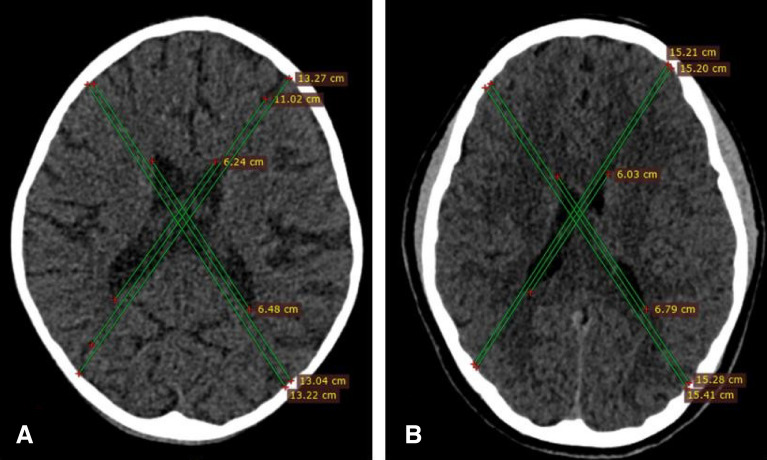
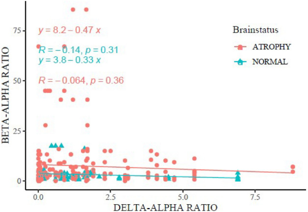
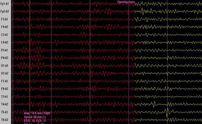
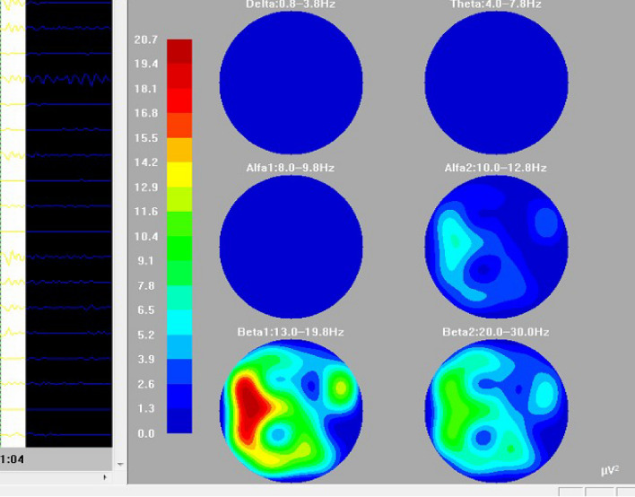
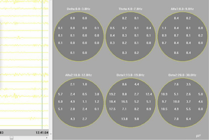
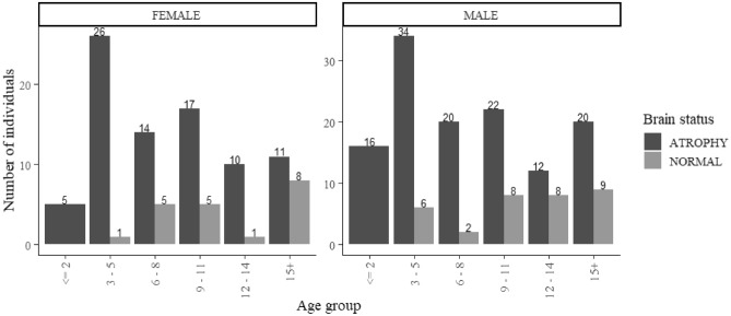
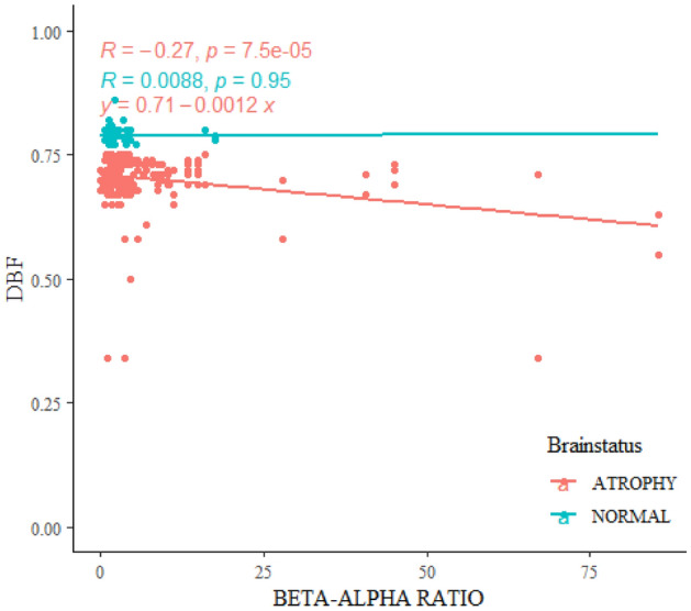
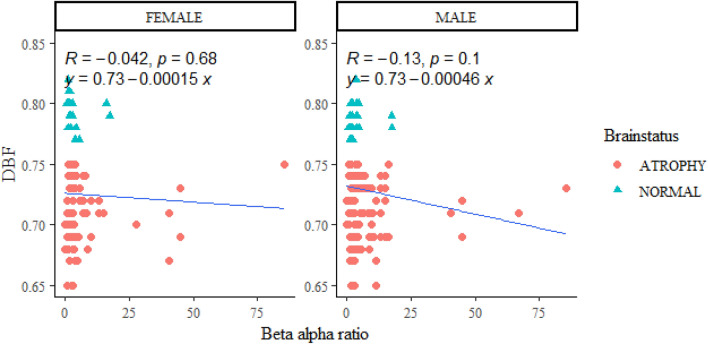
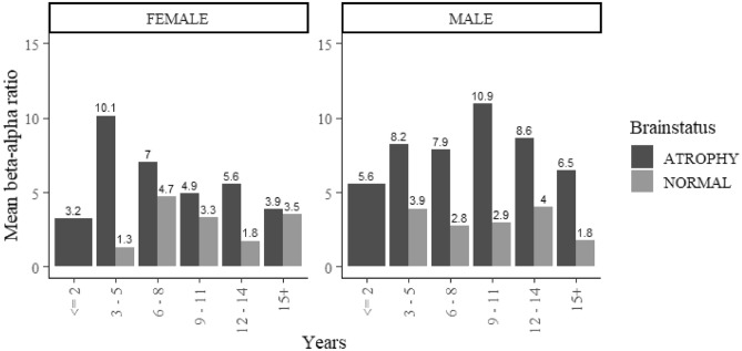
Although it is a normal involution process in advanced age, brain atrophy-also termed atrophic encephalopathy-can also occur prematurely in childhood as a consequential effect of brain tissues injury through trauma or central nervous system infection, though in both normal and premature occurrences this condition always presents with loss of volume relative to the skull. A common tool for the functional study of brain activities is an electroencephalogram, but analyses of this have reportedly identified mismatches between qualitative and quantitative forms, particularly in the use of Delta-alpha ratio (DAR) indices, meaning that the values may be case dependent. The current study thus examines the value of Focused Occipital Beta-Alpha Ratio (FOBAR) as a modified biomarker for evaluating brain functional changes resulting from brain atrophy. This cross-sectional design study involves 260 patients under 18 years of age. Specifically, 207 patients with brain atrophy are compared with 53 control subjects with CT scan-proven normal brain volume. All the children underwent digital electroencephalography with brain mapping. Results show that alpha posterior dominant rhythm was present in 88 atrophic children and 44 controls. Beta as posterior dominant rhythm was present in an overwhelming 91.5% of atrophic subjects, with 0.009 p-values. The focused occipital Beta-alpha ratio correlated significantly with brain volume loss presented in diagonal brain fraction. The FOBAR and DAR values of the QEEG showed no significant correlation. This work concludes that QEEG cerebral dysfunctional studies may be etiologically and case dependent from the nature of the brain injury. Also, the focused Beta-alpha ratio of the QEEG is a prospective and potential biomarker of consideration in studying childhood atrophic encephalopathy.

摘要

虽然脑萎缩-也称为萎缩性脑病-是老年人的正常退行性过程,但在儿童时期,由于创伤或中枢神经系统感染导致脑组织损伤,也可能会提前发生。尽管在正常和提前发生的情况下,这种情况总是表现为相对于颅骨的体积损失。一种用于研究大脑活动的常见功能工具是脑电图,但据报道,对其分析发现了定性和定量形式之间的不匹配,特别是在使用 Delta-alpha 比(DAR)指数方面,这意味着这些值可能取决于具体情况。因此,本研究检查了聚焦枕部 Beta-Alpha 比(FOBAR)作为评估脑萎缩引起的大脑功能变化的改良生物标志物的价值。这项横断面设计研究涉及 260 名 18 岁以下的患者。具体来说,将 207 名脑萎缩患者与经 CT 扫描证实大脑体积正常的 53 名对照受试者进行比较。所有儿童均接受数字脑电图和脑图检查。结果显示,88 名萎缩儿童和 44 名对照者存在 alpha 后优势节律。91.5%的萎缩患者存在压倒性的 Beta 后优势节律,p 值为 0.009。焦点枕部 Beta-alpha 比与对角脑分数中表现出的脑体积损失显著相关。QEEG 的 FOBAR 和 DAR 值无显著相关性。本研究得出结论,QEEG 脑功能障碍研究可能与脑损伤的病因和病例有关。此外,QEEG 的焦点 Beta-alpha 比是研究儿童萎缩性脑病的潜在生物标志物。

https://cdn.ncbi.nlm.nih.gov/pmc/blobs/3911/9352776/dde218edd0bb/41598_2022_17062_Fig9_HTML.jpg
https://cdn.ncbi.nlm.nih.gov/pmc/blobs/3911/9352776/dc6de6ea5565/41598_2022_17062_Fig1_HTML.jpg
https://cdn.ncbi.nlm.nih.gov/pmc/blobs/3911/9352776/f0b73da76152/41598_2022_17062_Fig2_HTML.jpg
https://cdn.ncbi.nlm.nih.gov/pmc/blobs/3911/9352776/a580d1392d5c/41598_2022_17062_Fig3_HTML.jpg
https://cdn.ncbi.nlm.nih.gov/pmc/blobs/3911/9352776/28aa699a5f43/41598_2022_17062_Fig4_HTML.jpg
https://cdn.ncbi.nlm.nih.gov/pmc/blobs/3911/9352776/ed9bd6829a9b/41598_2022_17062_Fig5_HTML.jpg
https://cdn.ncbi.nlm.nih.gov/pmc/blobs/3911/9352776/b73e60824659/41598_2022_17062_Fig6_HTML.jpg
https://cdn.ncbi.nlm.nih.gov/pmc/blobs/3911/9352776/040edc1d4f20/41598_2022_17062_Fig7_HTML.jpg
https://cdn.ncbi.nlm.nih.gov/pmc/blobs/3911/9352776/6c3d9b6d2196/41598_2022_17062_Fig8_HTML.jpg
https://cdn.ncbi.nlm.nih.gov/pmc/blobs/3911/9352776/dde218edd0bb/41598_2022_17062_Fig9_HTML.jpg

相似文献

1
The focused quantitative EEG bio-marker in studying childhood atrophic encephalopathy.研究儿童脑萎缩性脑病的聚焦定量 EEG 生物标志物。
Sci Rep. 2022 Aug 4;12(1):13437. doi: 10.1038/s41598-022-17062-w.
2
Quantitative spectral analysis of EEG in psychiatry revisited: drawing signs out of numbers in a clinical setting.精神病学中脑电图定量光谱分析的再探讨:在临床环境中从数字中提取特征。
Clin Neurophysiol. 2003 Dec;114(12):2294-306. doi: 10.1016/s1388-2457(03)00228-1.
3
Frontal EEG delta/alpha ratio and screening for post-stroke cognitive deficits: the power of four electrodes.额叶脑电图δ/α比值与中风后认知缺陷筛查:四个电极的作用
Int J Psychophysiol. 2014 Oct;94(1):19-24. doi: 10.1016/j.ijpsycho.2014.06.012. Epub 2014 Jun 24.
4
Spectral Electroencephalogram Analysis for the Evaluation of Encephalopathy Grade in Children With Acute Liver Failure.用于评估急性肝衰竭患儿肝性脑病分级的频谱脑电图分析
Pediatr Crit Care Med. 2017 Jan;18(1):64-72. doi: 10.1097/PCC.0000000000001016.
5
Quantitative EEG as a predictive biomarker for Parkinson disease dementia.定量脑电图作为帕金森病痴呆的预测生物标志物。
Neurology. 2011 Jul 12;77(2):118-24. doi: 10.1212/WNL.0b013e318224af8d. Epub 2011 Jun 1.
6
Defining abnormal slow EEG activity in acute ischaemic stroke: Delta/alpha ratio as an optimal QEEG index.急性缺血性卒中异常慢脑电图活动的定义:δ/α比值作为最佳定量脑电图指标
Clin Neurophysiol. 2016 Feb;127(2):1452-1459. doi: 10.1016/j.clinph.2015.07.014. Epub 2015 Jul 22.
7
Quantitative EEG and dynamic susceptibility contrast MRI in Alzheimer's disease: a correlative study.阿尔茨海默病的定量脑电图与动态磁敏感对比磁共振成像:一项相关性研究。
Clin Neurophysiol. 2003 Jul;114(7):1210-6. doi: 10.1016/s1388-2457(03)00085-3.
8
Clinical correlates of quantitative EEG alterations in alcoholic patients.酒精性患者定量脑电图改变的临床相关性
Clin Neurophysiol. 2006 Apr;117(4):740-51. doi: 10.1016/j.clinph.2005.12.021. Epub 2006 Feb 21.
9
Hippocampal atrophy and EEG markers in subjects with mild cognitive impairment.轻度认知障碍患者的海马萎缩与脑电图标志物
Clin Neurophysiol. 2007 Dec;118(12):2716-29. doi: 10.1016/j.clinph.2007.09.059. Epub 2007 Oct 30.
10
Comparative analysis of background EEG activity based on MRI findings in neonatal hypoxic-ischemic encephalopathy: a standardized, low-resolution, brain electromagnetic tomography (sLORETA) study.基于 MRI 发现的新生儿缺氧缺血性脑病背景 EEG 活动的比较分析:一项标准化、低分辨率、脑电磁断层扫描(sLORETA)研究。
BMC Neurol. 2022 Jun 3;22(1):204. doi: 10.1186/s12883-022-02736-9.

本文引用的文献

1
Alpha/Theta Ratio Neurofeedback Training for Attention Enhancement in Normal Developing Children: A Brief Report.α/θ比率神经反馈训练对正常发育儿童注意力增强的作用:简要报告
Appl Psychophysiol Biofeedback. 2022 Sep;47(3):223-229. doi: 10.1007/s10484-022-09550-6. Epub 2022 Jun 13.
2
Progressive cerebral atrophies in three children with COL4A1 mutations.三名患有COL4A1基因突变儿童的进行性脑萎缩
Brain Dev. 2021 Nov;43(10):1033-1038. doi: 10.1016/j.braindev.2021.06.008. Epub 2021 Jul 17.
3
Using EEG Alpha States to Understand Learning During Alpha Neurofeedback Training for Chronic Pain.
利用脑电图α波状态理解慢性疼痛的α波神经反馈训练期间的学习情况。
Front Neurosci. 2021 Feb 22;14:620666. doi: 10.3389/fnins.2020.620666. eCollection 2020.
4
Quantification of stroke lesion volume using epidural EEG in a cerebral ischaemic rat model.硬膜外 EEG 在脑缺血大鼠模型中对中风病灶体积的定量评估。
Sci Rep. 2021 Jan 27;11(1):2308. doi: 10.1038/s41598-021-81912-2.
5
Novel multi-linear quantitative brain volume formula for manual radiological evaluation of brain atrophy.用于脑萎缩手动放射学评估的新型多线性定量脑容量公式。
Eur J Radiol Open. 2020 Nov 13;7:100281. doi: 10.1016/j.ejro.2020.100281. eCollection 2020.
6
The neuroimaging magnitude of pediatric brain atrophy in northern Tanzania.坦桑尼亚北部小儿脑萎缩的神经影像学程度
Pan Afr Med J. 2020 May 21;36:25. doi: 10.11604/pamj.2020.36.25.22515. eCollection 2020.
7
Dynamic Regional Brain Atrophy Rates in the First Year After Ischemic Stroke.缺血性脑卒中后第一年的动态区域性脑萎缩率。
Stroke. 2020 Sep;51(9):e183-e192. doi: 10.1161/STROKEAHA.120.030256. Epub 2020 Aug 10.
8
CT-Based Classification of Acute Cerebral Edema: Association with Intracranial Pressure and Outcome.基于 CT 的急性脑水肿分类:与颅内压和预后的关系。
J Neuroimaging. 2020 Sep;30(5):640-647. doi: 10.1111/jon.12736. Epub 2020 May 28.
9
Real-time fMRI for brain-computer interfacing.用于脑机接口的实时功能磁共振成像
Handb Clin Neurol. 2020;168:289-302. doi: 10.1016/B978-0-444-63934-9.00021-4.
10
Alpha Synchrony and the Neurofeedback Control of Spatial Attention.阿尔法同步与空间注意的神经反馈控制。
Neuron. 2020 Feb 5;105(3):577-587.e5. doi: 10.1016/j.neuron.2019.11.001. Epub 2019 Dec 4.