Center for Cell Death, Injury & Regeneration, Medical University of South Carolina, Charleston, South Carolina, USA; Department of Drug Discovery & Biomedical Sciences, Medical University of South Carolina, Charleston, South Carolina, USA.
Department of Drug Discovery & Biomedical Sciences, Medical University of South Carolina, Charleston, South Carolina, USA.
J Biol Chem. 2022 Sep;298(9):102336. doi: 10.1016/j.jbc.2022.102336. Epub 2022 Aug 2.
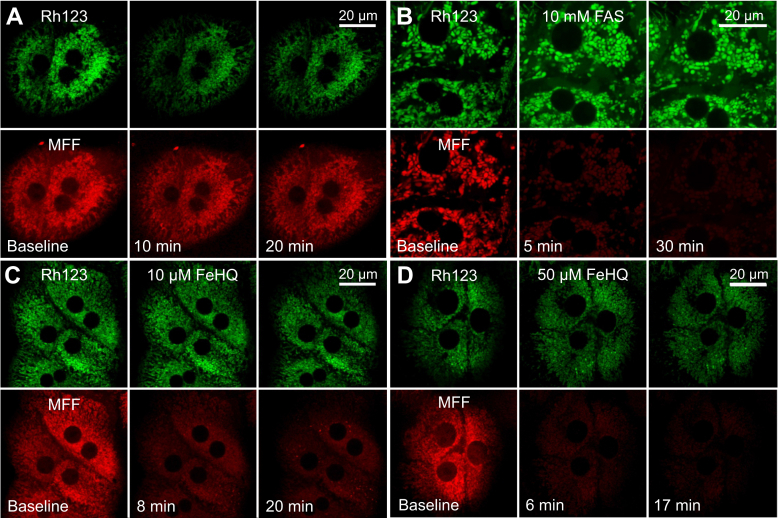
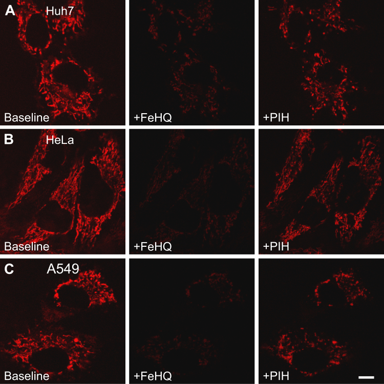
Mitochondrial chelatable iron contributes to the severity of several injury processes, including ischemia/reperfusion, oxidative stress, and drug toxicity. However, methods to measure this species in living cells are lacking. To measure mitochondrial chelatable iron in living cells, here we synthesized a new fluorescent indicator, mitoferrofluor (MFF). We designed cationic MFF to accumulate electrophoretically in polarized mitochondria, where a reactive group then forms covalent adducts with mitochondrial proteins to retain MFF even after subsequent depolarization. We also show in cell-free medium that Fe (and Cu), but not Fe, Ca, or other biologically relevant divalent cations, strongly quenched MFF fluorescence. Using confocal microscopy, we demonstrate in hepatocytes that red MFF fluorescence colocalized with the green fluorescence of the mitochondrial membrane potential (ΔΨ) indicator, rhodamine 123 (Rh123), indicating selective accumulation into the mitochondria. Unlike Rh123, mitochondria retained MFF after ΔΨ collapse. Furthermore, intracellular delivery of iron with membrane-permeant Fe/8-hydroxyquinoline (FeHQ) quenched MFF fluorescence by ∼80% in hepatocytes and other cell lines, which was substantially restored by the membrane-permeant transition metal chelator pyridoxal isonicotinoyl hydrazone. We also show FeHQ quenched the fluorescence of cytosolically coloaded calcein, another Fe indicator, confirming that Fe in FeHQ undergoes intracellular reduction to Fe. Finally, MFF fluorescence did not change after addition of the calcium mobilizer thapsigargin, which shows MFF is insensitive to physiologically relevant increases of mitochondrial Ca. In conclusion, the new sensor reagent MFF fluorescence is an indicator of mitochondrial chelatable Fe in normal hepatocytes with polarized mitochondria as well as in cells undergoing loss of ΔΨ.
线粒体可螯合铁有助于几种损伤过程的严重程度,包括缺血/再灌注、氧化应激和药物毒性。然而,缺乏测量这种物质在活细胞中的方法。为了测量活细胞中线粒体可螯合铁,我们在这里合成了一种新的荧光指示剂,mitoferrofluor(MFF)。我们设计了阳离子 MFF,使其在极化的线粒体中通过电泳积累,然后在反应基团与线粒体蛋白形成共价加合物,即使在随后的去极化后,MFF 也能保留下来。我们还在无细胞介质中表明,Fe(和 Cu),而不是 Fe、Ca 或其他生物相关的二价阳离子,强烈猝灭 MFF 荧光。使用共聚焦显微镜,我们在肝细胞中证明红色 MFF 荧光与线粒体膜电位(ΔΨ)指示剂 rhodamine 123(Rh123)的绿色荧光共定位,表明其选择性地积累到线粒体中。与 Rh123 不同,线粒体在 ΔΨ 崩溃后仍保留 MFF。此外,用膜透性铁/8-羟基喹啉(FeHQ)将铁递送到细胞内,使肝细胞和其他细胞系中的 MFF 荧光猝灭约 80%,而用膜透性过渡金属螯合剂吡啶甲酰异羟肟酸显著恢复。我们还表明,FeHQ 猝灭细胞质共加载的 calcein(另一种 Fe 指示剂)的荧光,证实 FeHQ 中的 Fe 在内质网还原为 Fe。最后,加入钙动员剂 thapsigargin 后 MFF 荧光没有变化,表明 MFF 对线粒体 Ca 的生理相关增加不敏感。总之,新的传感器试剂 MFF 荧光是正常具有极化线粒体的肝细胞以及经历 ΔΨ 丧失的细胞中线粒体可螯合铁的指示剂。