• 文献检索
  • 文档翻译
  • 深度研究
  • 学术资讯
  • Suppr Zotero 插件Zotero 插件
  • 邀请有礼
  • 套餐&价格
  • 历史记录
应用&插件
Suppr Zotero 插件Zotero 插件浏览器插件Mac 客户端Windows 客户端微信小程序
定价
高级版会员购买积分包购买API积分包
服务
文献检索文档翻译深度研究API 文档MCP 服务
关于我们
关于 Suppr公司介绍联系我们用户协议隐私条款
关注我们

Suppr 超能文献

核心技术专利:CN118964589B侵权必究
粤ICP备2023148730 号-1Suppr @ 2026

文献检索

告别复杂PubMed语法,用中文像聊天一样搜索,搜遍4000万医学文献。AI智能推荐,让科研检索更轻松。

立即免费搜索

文件翻译

保留排版,准确专业,支持PDF/Word/PPT等文件格式,支持 12+语言互译。

免费翻译文档

深度研究

AI帮你快速写综述,25分钟生成高质量综述,智能提取关键信息,辅助科研写作。

立即免费体验

基于动态对比增强磁共振成像的深度学习模型能够准确预测乳腺病变的良恶性。

A deep learning model based on dynamic contrast-enhanced magnetic resonance imaging enables accurate prediction of benign and malignant breast lessons.

作者信息

Chen Yanhong, Wang Lijun, Luo Ran, Wang Shuang, Wang Heng, Gao Fei, Wang Dengbin

机构信息

Department of Radiology, Xinhua Hospital Affiliated to Shanghai Jiao Tong University School of Medicine, Shanghai, China.

Department of Medicine, Beijing Medicinovo Technology Co., Ltd., Beijing, China.

出版信息

Front Oncol. 2022 Jul 22;12:943415. doi: 10.3389/fonc.2022.943415. eCollection 2022.

DOI:10.3389/fonc.2022.943415
PMID:35936673
原文链接:https://pmc.ncbi.nlm.nih.gov/articles/PMC9353744/
Abstract

OBJECTIVES

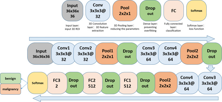
The study aims to investigate the value of a convolutional neural network (CNN) based on dynamic contrast-enhanced magnetic resonance imaging (DCE-MRI) in predicting malignancy of breast lesions.

METHODS

We developed a CNN model based on DCE-MRI to characterize breast lesions. Between November 2018 and October 2019, 6,165 slices of 364 lesions (234 malignant, 130 benign) in 364 patients were pooled in the training/validation set. Lesions were semi-automatically segmented by two breast radiologists using ITK-SNAP software. The standard of reference was histologic consequences. Algorithm performance was evaluated in an independent testing set of 1,560 slices of 127 lesions in 127 patients using weighted sums of the area under the curve (AUC) scores.

RESULTS

The area under the receiver operating characteristic (ROC) curve was 0.955 for breast cancer prediction while the accuracy, sensitivity, and specificity were 90.3, 96.2, and 79.0%, respectively, in the slice-based method. In the case-based method, the efficiency of the model changed by adjusting the standard for the number of positive slices. When a lesion with three or more positive slices was determined as malignant, the sensitivity was above 90%, with a specificity of nearly 60% and an accuracy higher than 80%.

CONCLUSION

The CNN model based on DCE-MRI demonstrated high accuracy for predicting malignancy among the breast lesions. This method should be validated in a larger and independent cohort.

摘要

目的

本研究旨在探讨基于动态对比增强磁共振成像(DCE-MRI)的卷积神经网络(CNN)在预测乳腺病变恶性程度方面的价值。

方法

我们开发了一种基于DCE-MRI的CNN模型来表征乳腺病变。在2018年11月至2019年10月期间,将364例患者的364个病变(234个恶性,130个良性)的6165层图像纳入训练/验证集。两名乳腺放射科医生使用ITK-SNAP软件对病变进行半自动分割。参考标准为组织学结果。使用曲线下面积(AUC)分数的加权总和,在127例患者的127个病变的1560层图像的独立测试集中评估算法性能。

结果

在基于切片的方法中,乳腺癌预测的受试者操作特征(ROC)曲线下面积为0.955,而准确率、敏感性和特异性分别为90.3%、96.2%和79.0%。在基于病例的方法中,通过调整阳性切片数量的标准来改变模型的效率。当将具有三个或更多阳性切片的病变判定为恶性时,敏感性高于90%,特异性接近60%,准确率高于80%。

结论

基于DCE-MRI的CNN模型在预测乳腺病变恶性程度方面显示出较高的准确性。该方法应在更大的独立队列中进行验证。

https://cdn.ncbi.nlm.nih.gov/pmc/blobs/db0b/9353744/71bb01eadfd1/fonc-12-943415-g008.jpg
https://cdn.ncbi.nlm.nih.gov/pmc/blobs/db0b/9353744/3da98ea47401/fonc-12-943415-g001.jpg
https://cdn.ncbi.nlm.nih.gov/pmc/blobs/db0b/9353744/760e2df5cbb5/fonc-12-943415-g002.jpg
https://cdn.ncbi.nlm.nih.gov/pmc/blobs/db0b/9353744/2f0d533eaf24/fonc-12-943415-g003.jpg
https://cdn.ncbi.nlm.nih.gov/pmc/blobs/db0b/9353744/dc053f055ea8/fonc-12-943415-g004.jpg
https://cdn.ncbi.nlm.nih.gov/pmc/blobs/db0b/9353744/afd772e928e7/fonc-12-943415-g005.jpg
https://cdn.ncbi.nlm.nih.gov/pmc/blobs/db0b/9353744/6672076befdd/fonc-12-943415-g006.jpg
https://cdn.ncbi.nlm.nih.gov/pmc/blobs/db0b/9353744/4d991f22686d/fonc-12-943415-g007.jpg
https://cdn.ncbi.nlm.nih.gov/pmc/blobs/db0b/9353744/71bb01eadfd1/fonc-12-943415-g008.jpg

相似文献

1
A deep learning model based on dynamic contrast-enhanced magnetic resonance imaging enables accurate prediction of benign and malignant breast lessons.基于动态对比增强磁共振成像的深度学习模型能够准确预测乳腺病变的良恶性。
Front Oncol. 2022 Jul 22;12:943415. doi: 10.3389/fonc.2022.943415. eCollection 2022.
2
Deep-learning approach with convolutional neural network for classification of maximum intensity projections of dynamic contrast-enhanced breast magnetic resonance imaging.基于卷积神经网络的深度学习方法在动态对比增强乳腺磁共振成像最大强度投影中的分类。
Magn Reson Imaging. 2021 Jan;75:1-8. doi: 10.1016/j.mri.2020.10.003. Epub 2020 Oct 10.
3
Automatic deep learning method for detection and classification of breast lesions in dynamic contrast-enhanced magnetic resonance imaging.动态对比增强磁共振成像中乳腺病变检测与分类的自动深度学习方法
Quant Imaging Med Surg. 2023 Apr 1;13(4):2620-2633. doi: 10.21037/qims-22-323. Epub 2023 Jan 14.
4
Diagnosis of Benign and Malignant Breast Lesions on DCE-MRI by Using Radiomics and Deep Learning With Consideration of Peritumor Tissue.基于瘤周组织考虑的放射组学和深度学习在 DCE-MRI 上对乳腺良恶性病变的诊断。
J Magn Reson Imaging. 2020 Mar;51(3):798-809. doi: 10.1002/jmri.26981. Epub 2019 Nov 1.
5
Detection and characterization of MRI breast lesions using deep learning.利用深度学习检测和描述 MRI 乳腺病变。
Diagn Interv Imaging. 2019 Apr;100(4):219-225. doi: 10.1016/j.diii.2019.02.008. Epub 2019 Mar 26.
6
Differentiation of breast lesions on dynamic contrast-enhanced magnetic resonance imaging (DCE-MRI) using deep transfer learning based on DenseNet201.基于 DenseNet201 的深度迁移学习在动态对比增强磁共振成像(DCE-MRI)上对乳腺病变的鉴别诊断。
Medicine (Baltimore). 2022 Nov 11;101(45):e31214. doi: 10.1097/MD.0000000000031214.
7
Independent validation of machine learning in diagnosing breast Cancer on magnetic resonance imaging within a single institution.在单家医疗机构内使用机器学习对乳腺磁共振成像进行独立验证诊断乳腺癌。
Cancer Imaging. 2019 Sep 18;19(1):64. doi: 10.1186/s40644-019-0252-2.
8
Multi-input deep learning architecture for predicting breast tumor response to chemotherapy using quantitative MR images.基于多输入深度学习架构的利用定量 MRI 预测乳腺癌对化疗的反应
Int J Comput Assist Radiol Surg. 2020 Sep;15(9):1491-1500. doi: 10.1007/s11548-020-02209-9. Epub 2020 Jun 16.
9
Transfer Learning Strategy Based on Unsupervised Learning and Ensemble Learning for Breast Cancer Molecular Subtype Prediction Using Dynamic Contrast-Enhanced MRI.基于无监督学习和集成学习的迁移学习策略用于使用动态对比增强磁共振成像预测乳腺癌分子亚型
J Magn Reson Imaging. 2022 May;55(5):1518-1534. doi: 10.1002/jmri.27955. Epub 2021 Oct 20.
10
Diagnostic efficiency of multi-modal MRI based deep learning with Sobel operator in differentiating benign and malignant breast mass lesions-a retrospective study.基于多模态MRI和Sobel算子的深度学习在鉴别乳腺良恶性肿块病变中的诊断效能——一项回顾性研究
PeerJ Comput Sci. 2023 Jul 17;9:e1460. doi: 10.7717/peerj-cs.1460. eCollection 2023.

引用本文的文献

1
Deep learning-based breast cancer diagnosis in breast MRI: systematic review and meta-analysis.基于深度学习的乳腺MRI乳腺癌诊断:系统评价与荟萃分析
Eur Radiol. 2025 Feb 5. doi: 10.1007/s00330-025-11406-6.
2
A twin convolutional neural network with hybrid binary optimizer for multimodal breast cancer digital image classification.一种具有混合二进制优化器的双卷积神经网络,用于多模态乳腺癌数字图像分类。
Sci Rep. 2024 Jan 6;14(1):692. doi: 10.1038/s41598-024-51329-8.
3
AI-Based Cancer Detection Model for Contrast-Enhanced Mammography.基于人工智能的对比增强乳腺X线摄影癌症检测模型

本文引用的文献

1
Diagnostic Performance of 2D and 3D T2WI-Based Radiomics Features With Machine Learning Algorithms to Distinguish Solid Solitary Pulmonary Lesion.基于二维和三维T2加权成像的影像组学特征联合机器学习算法鉴别实性孤立性肺结节的诊断效能
Front Oncol. 2021 Nov 18;11:683587. doi: 10.3389/fonc.2021.683587. eCollection 2021.
2
Multi-Class brain normality and abnormality diagnosis using modified Faster R-CNN.使用改进的Faster R-CNN进行多类别脑正常与异常诊断。
Int J Med Inform. 2021 Nov;155:104576. doi: 10.1016/j.ijmedinf.2021.104576. Epub 2021 Sep 16.
3
Global Cancer Statistics 2020: GLOBOCAN Estimates of Incidence and Mortality Worldwide for 36 Cancers in 185 Countries.
Bioengineering (Basel). 2023 Aug 17;10(8):974. doi: 10.3390/bioengineering10080974.
《全球癌症统计数据 2020:全球 185 个国家和地区 36 种癌症的发病率和死亡率估计》。
CA Cancer J Clin. 2021 May;71(3):209-249. doi: 10.3322/caac.21660. Epub 2021 Feb 4.
4
A deep learning model integrating mammography and clinical factors facilitates the malignancy prediction of BI-RADS 4 microcalcifications in breast cancer screening.深度学习模型整合乳腺 X 线摄影和临床因素有助于预测乳腺癌筛查中 BI-RADS 4 微钙化的恶性程度。
Eur Radiol. 2021 Aug;31(8):5902-5912. doi: 10.1007/s00330-020-07659-y. Epub 2021 Jan 26.
5
Lung Nodule Detection based on Faster R-CNN Framework.基于Faster R-CNN框架的肺结节检测
Comput Methods Programs Biomed. 2021 Mar;200:105866. doi: 10.1016/j.cmpb.2020.105866. Epub 2020 Nov 22.
6
Key steps for effective breast cancer prevention.有效预防乳腺癌的关键步骤。
Nat Rev Cancer. 2020 Aug;20(8):417-436. doi: 10.1038/s41568-020-0266-x. Epub 2020 Jun 11.
7
International evaluation of an AI system for breast cancer screening.国际乳腺癌筛查人工智能系统评估。
Nature. 2020 Jan;577(7788):89-94. doi: 10.1038/s41586-019-1799-6. Epub 2020 Jan 1.
8
Ultrasonic Diagnosis of Breast Nodules Using Modified Faster R-CNN.基于改进的 Faster R-CNN 的乳腺结节超声诊断
Ultrason Imaging. 2019 Nov;41(6):353-367. doi: 10.1177/0161734619882683.
9
Artificial Intelligence for Mammography and Digital Breast Tomosynthesis: Current Concepts and Future Perspectives.人工智能在乳腺 X 线摄影和数字乳腺断层合成中的应用:现状与未来展望。
Radiology. 2019 Nov;293(2):246-259. doi: 10.1148/radiol.2019182627. Epub 2019 Sep 24.
10
A Deep Learning Model to Triage Screening Mammograms: A Simulation Study.深度学习模型在乳房 X 光筛查中的分诊作用:一项模拟研究。
Radiology. 2019 Oct;293(1):38-46. doi: 10.1148/radiol.2019182908. Epub 2019 Aug 6.