Section on Quantitative Imaging and Tissue Sciences, Eunice Kennedy Shriver National Institute of Child Health and Human Development, NIH, Bethesda, MD 20891, USA.
Multiscale Imaging and Integrative Biophysics Unit, Laboratory of Behavioral Neuroscience, National Institute on Aging, NIH, Baltimore, MD 21224, USA.
Brain. 2023 Mar 1;146(3):1212-1226. doi: 10.1093/brain/awac298.
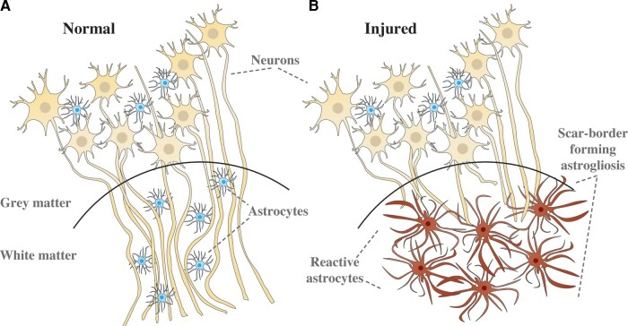
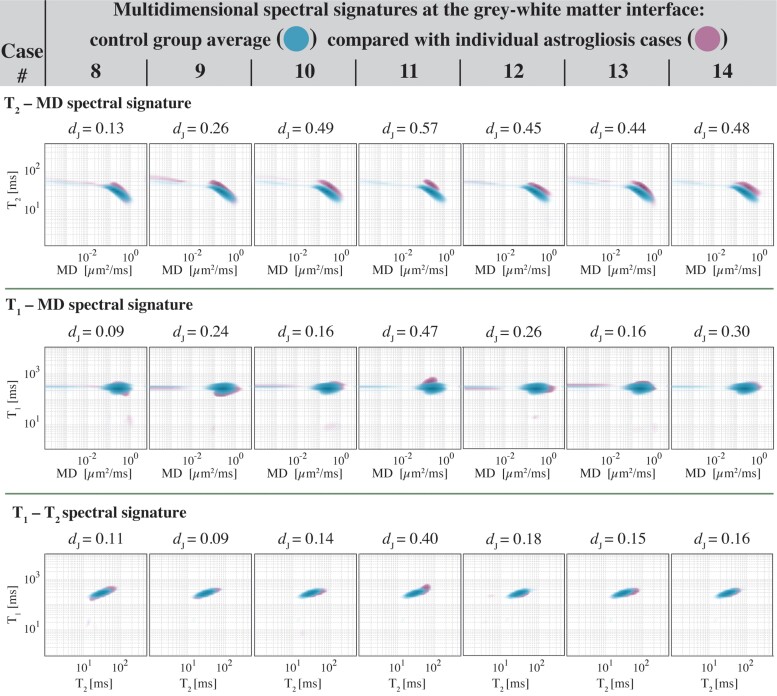
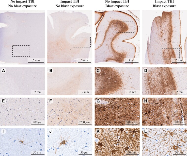
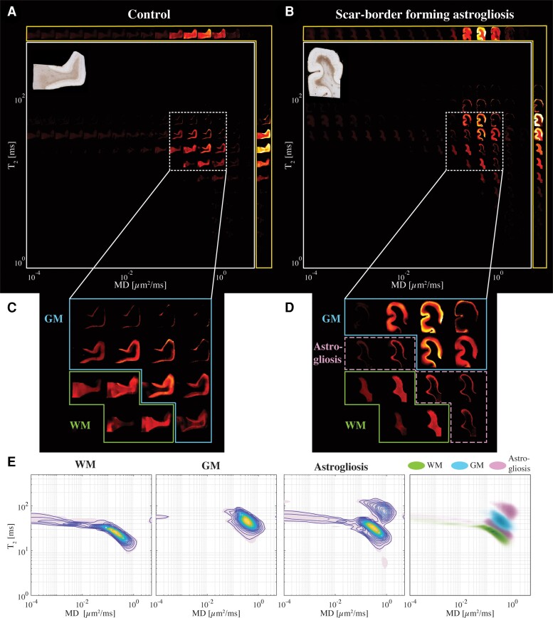
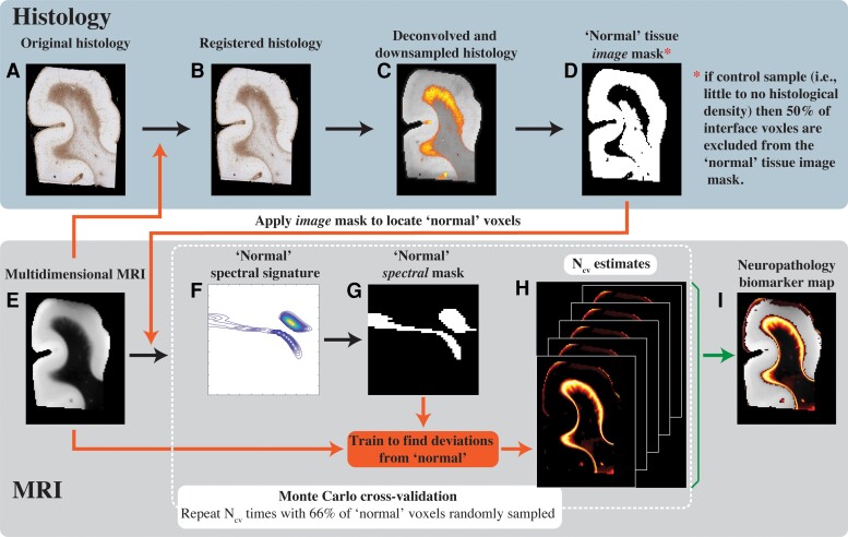
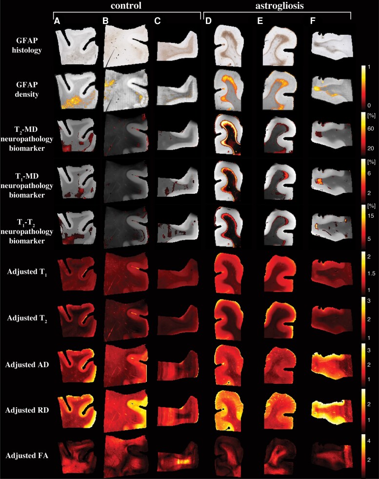
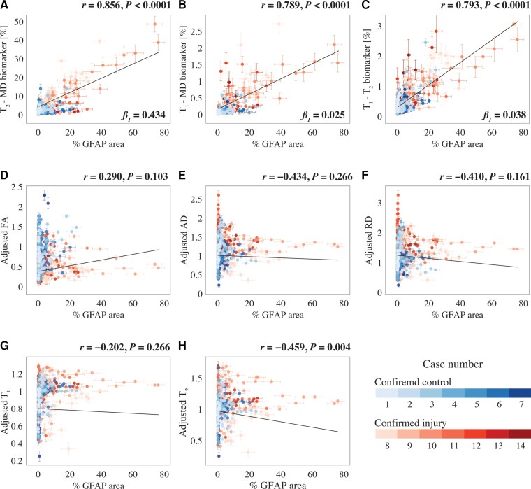
There are currently no non-invasive imaging methods available for astrogliosis assessment or mapping in the central nervous system despite its essential role in the response to many disease states, such as infarcts, neurodegenerative conditions, traumatic brain injury and infection. Multidimensional MRI is an increasingly employed imaging modality that maximizes the amount of encoded chemical and microstructural information by probing relaxation (T1 and T2) and diffusion mechanisms simultaneously. Here, we harness the exquisite sensitivity of this imagining modality to derive a signature of astrogliosis and disentangle it from normative brain at the individual level using machine learning. We investigated ex vivo cerebral cortical tissue specimens derived from seven subjects who sustained blast-induced injuries, which resulted in scar-border forming astrogliosis without being accompanied by other types of neuropathological abnormality, and from seven control brain donors. By performing a combined post-mortem radiology and histopathology correlation study we found that astrogliosis induces microstructural and chemical changes that are robustly detected with multidimensional MRI, and which can be attributed to astrogliosis because no axonal damage, demyelination or tauopathy were histologically observed in any of the cases in the study. Importantly, we showed that no one-dimensional T1, T2 or diffusion MRI measurement can disentangle the microscopic alterations caused by this neuropathology. Based on these findings, we developed a within-subject anomaly detection procedure that generates MRI-based astrogliosis biomarker maps ex vivo, which were significantly and strongly correlated with co-registered histological images of increased glial fibrillary acidic protein deposition (r = 0.856, P < 0.0001; r = 0.789, P < 0.0001; r = 0.793, P < 0.0001, for diffusion-T2, diffusion-T1 and T1-T2 multidimensional data sets, respectively). Our findings elucidate the underpinning of MRI signal response from astrogliosis, and the demonstrated high spatial sensitivity and specificity in detecting reactive astrocytes at the individual level, and if reproduced in vivo, will significantly impact neuroimaging studies of injury, disease, repair and aging, in which astrogliosis has so far been an invisible process radiologically.
目前,尽管星形胶质细胞在许多疾病状态(如梗死、神经退行性疾病、创伤性脑损伤和感染)的反应中起着至关重要的作用,但仍没有非侵入性的成像方法可用于星形胶质细胞的评估或定位。多维磁共振成像(MRI)是一种越来越被采用的成像方式,它通过同时探测弛豫(T1 和 T2)和扩散机制,最大限度地提高了编码的化学和微观结构信息的数量。在这里,我们利用这种成像方式的卓越敏感性,通过机器学习从个体水平上获得星形胶质细胞的特征,并将其与正常大脑区分开来。我们研究了来自 7 名因爆炸伤导致瘢痕边界形成星形胶质细胞增生但没有其他类型神经病理学异常的受试者和 7 名对照脑供体的离体大脑皮质组织标本。通过进行死后放射学和组织病理学相关性研究,我们发现星形胶质细胞增生诱导的微观结构和化学变化可以通过多维 MRI 进行稳健检测,并且可以归因于星形胶质细胞增生,因为在研究中的任何病例中都没有观察到轴突损伤、脱髓鞘或 tau 病的组织学表现。重要的是,我们表明,没有一种一维 T1、T2 或扩散 MRI 测量可以区分这种神经病理学引起的微观改变。基于这些发现,我们开发了一种基于个体的异常检测程序,该程序可以在离体状态下生成基于 MRI 的星形胶质细胞生物标志物图,这些图与共配准的胶质纤维酸性蛋白沉积增加的组织学图像显著且强烈相关(r = 0.856,P < 0.0001;r = 0.789,P < 0.0001;r = 0.793,P < 0.0001,分别为扩散-T2、扩散-T1 和 T1-T2 多维数据集)。我们的研究结果阐明了 MRI 信号响应与星形胶质细胞增生的关系,并证明了在个体水平上检测反应性星形胶质细胞的高空间灵敏度和特异性,如果在体内得到复制,将对损伤、疾病、修复和衰老的神经影像学研究产生重大影响,迄今为止,星形胶质细胞增生在影像学上一直是一个不可见的过程。