Benjamini Dan, Bouhrara Mustapha, Komlosh Michal E, Iacono Diego, Perl Daniel P, Brody David L, Basser Peter J
Section on Quantitative Imaging and Tissue Sciences, National Institute of Child Health and Human Development, National Institutes of Health, Bethesda, MD, United States.
Center for Neuroscience and Regenerative Medicine, Uniformed Services University of the Health Sciences, Bethesda, MD, United States.
Front Phys. 2021;9. doi: 10.3389/fphy.2021.737374. Epub 2021 Sep 16.
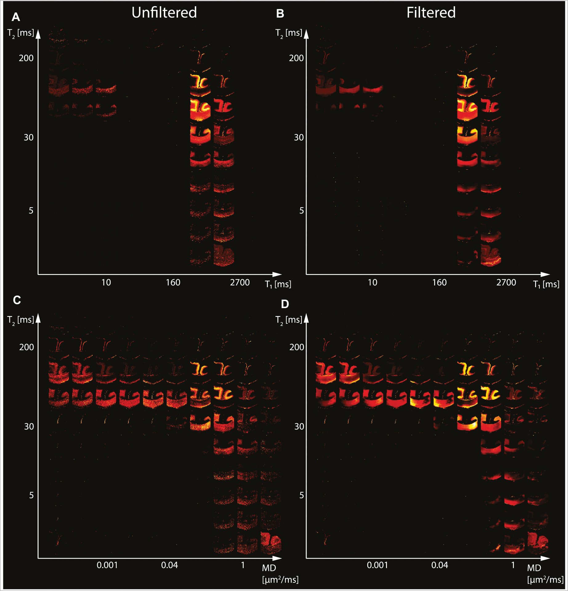
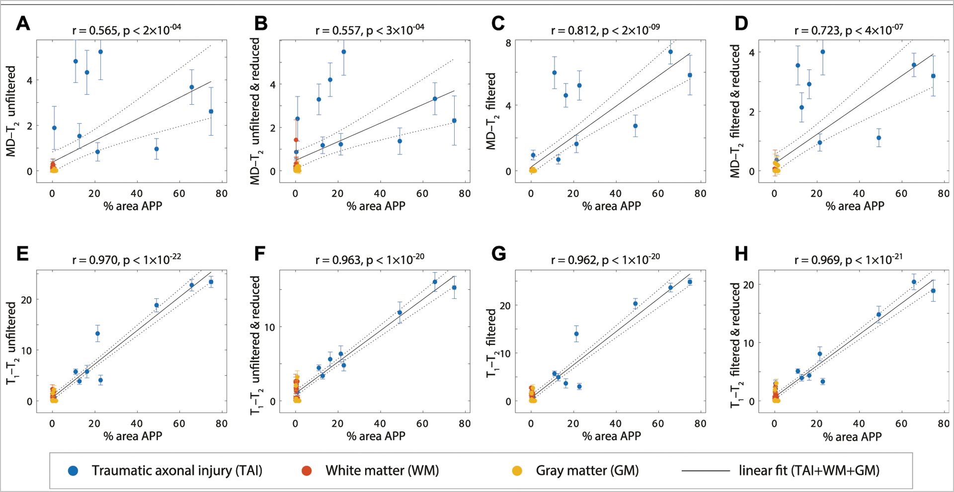
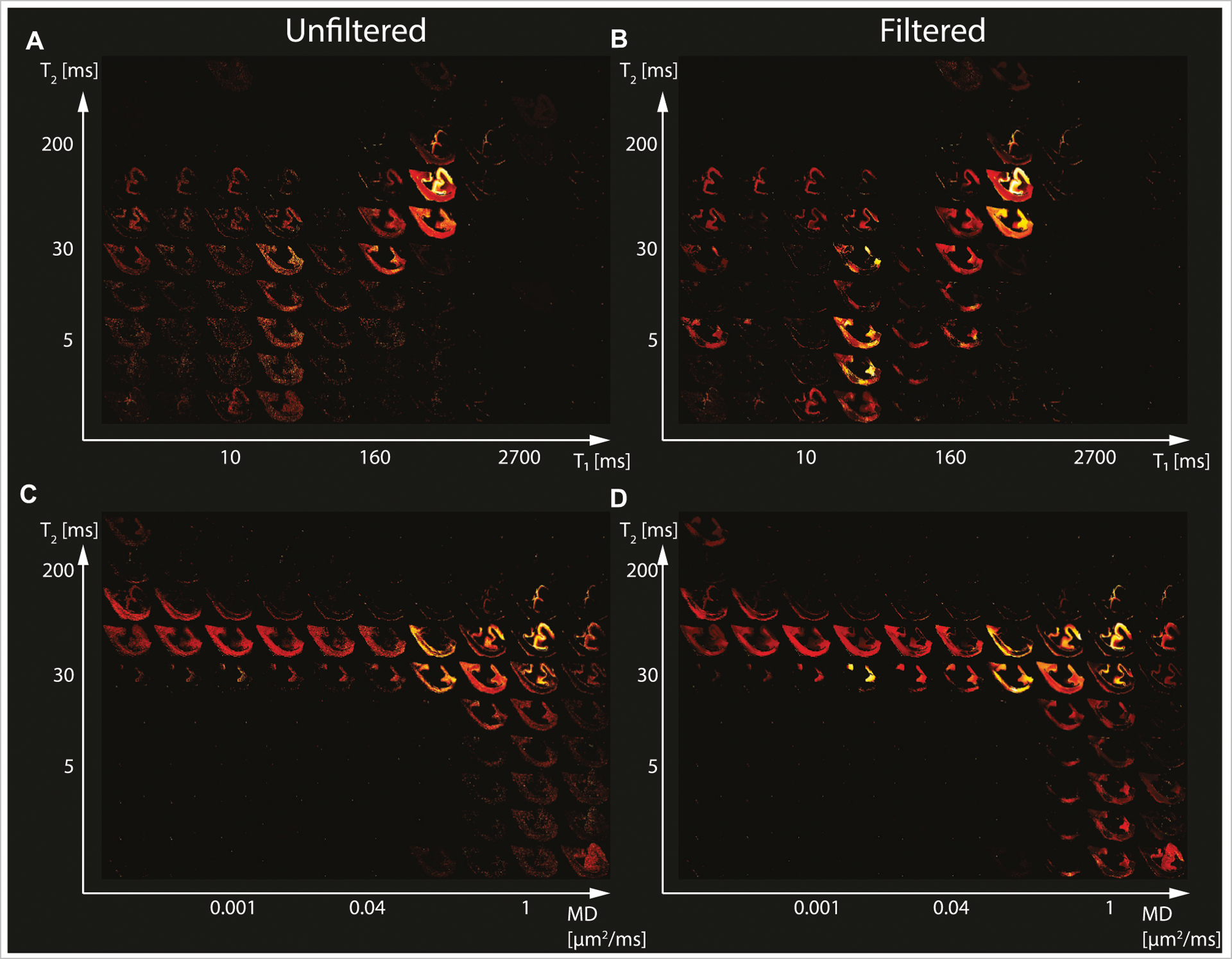
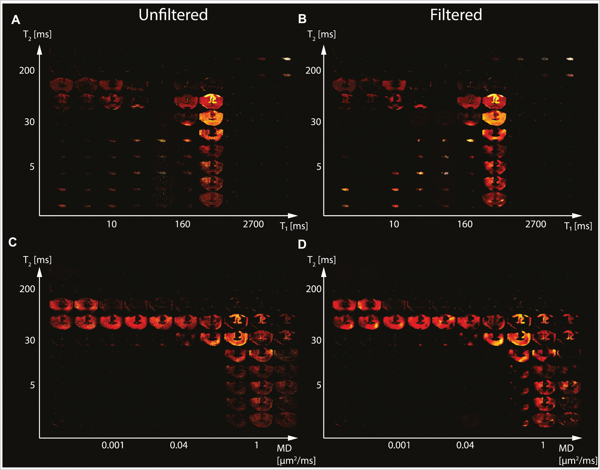
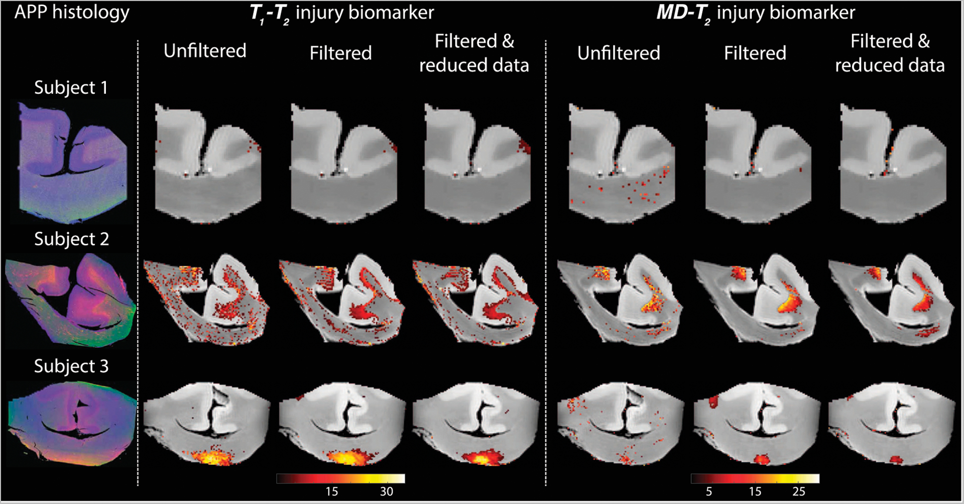
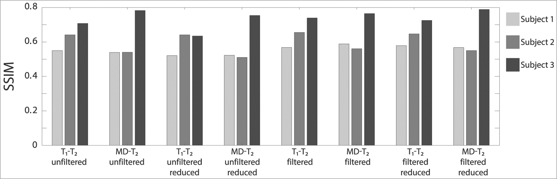
Multidimensional MRI is an emerging approach that simultaneously encodes water relaxation ( and ) and mobility (diffusion) and replaces voxel-averaged values with subvoxel distributions of those MR properties. While conventional (i.e., voxel-averaged) MRI methods cannot adequately quantify the microscopic heterogeneity of biological tissue, using subvoxel information allows to selectively map a specific --diffusion spectral range that corresponds to a group of tissue elements. The major obstacle to the adoption of rich, multidimensional MRI protocols for diagnostic or monitoring purposes is the prolonged scan time. Our main goal in the present study is to evaluate the performance of a nonlocal estimation of multispectral magnitudes (NESMA) filter on reduced datasets to limit the total acquisition time required for reliable multidimensional MRI characterization of the brain. Here we focused and reprocessed results from a recent study that identified potential imaging biomarkers of axonal injury pathology from the joint analysis of multidimensional MRI, in particular voxelwise - and diffusion- spectra in human Corpus Callosum, and histopathological data. We tested the performance of NESMA and its effect on the accuracy of the injury biomarker maps, relative to the co-registered histological reference. Noise reduction improved the accuracy of the resulting injury biomarker maps, while permitting data reduction of 35.7 and 59.6% from the full dataset for - and diffusion- cases, respectively. As successful clinical proof-of-concept applications of multidimensional MRI are continuously being introduced, reliable and robust noise removal and consequent acquisition acceleration would advance the field towards clinically-feasible diagnostic multidimensional MRI protocols.
多维磁共振成像(MRI)是一种新兴方法,它同时对水弛豫(和)及流动性(扩散)进行编码,并用这些磁共振特性的亚体素分布取代体素平均值。虽然传统的(即体素平均的)MRI方法无法充分量化生物组织的微观异质性,但使用亚体素信息可以选择性地绘制对应于一组组织成分的特定扩散光谱范围。采用丰富的多维MRI协议用于诊断或监测目的的主要障碍是扫描时间延长。我们在本研究中的主要目标是评估多光谱幅度非局部估计(NESMA)滤波器在简化数据集上的性能,以限制对大脑进行可靠的多维MRI表征所需的总采集时间。在此,我们重点关注并重新处理了一项近期研究的结果,该研究通过对多维MRI(特别是人胼胝体的体素逐点和扩散光谱)与组织病理学数据的联合分析,确定了轴突损伤病理的潜在成像生物标志物。我们测试了NESMA的性能及其对损伤生物标志物图谱准确性的影响,并与共同配准的组织学参考进行比较。降噪提高了所得损伤生物标志物图谱的准确性,同时在和扩散情况下分别允许从完整数据集中减少35.7%和59.6%的数据。随着多维MRI成功的临床概念验证应用不断推出,可靠且强大的噪声去除以及随之而来的采集加速将推动该领域朝着临床可行的诊断多维MRI协议发展。