Department of Endodontics and Periodontics, College of Stomatology, Dalian Medical University, 9 West Section, Lvshun South Road, Dalian, 116044, Liaoning Province, China.
Department of Pediatric Stomatology, The Third People's Hospital of Puyang City, Puyang, Henan Province, China.
BMC Oral Health. 2022 Aug 11;22(1):345. doi: 10.1186/s12903-022-02364-2.
Phosphoinositide 3-kinase (PI3K) is located within cells, and is involved in regulating cell survival, proliferation, apoptosis and angiogenesis. The purpose of this study was to investigate the role of PI3K in the process of bone destruction in apical periodontitis, and provide reference data for the treatment of this disease.
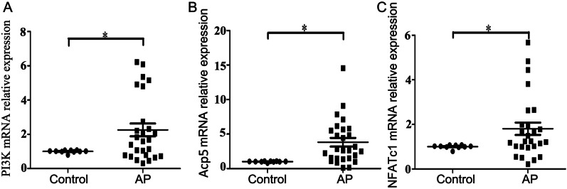
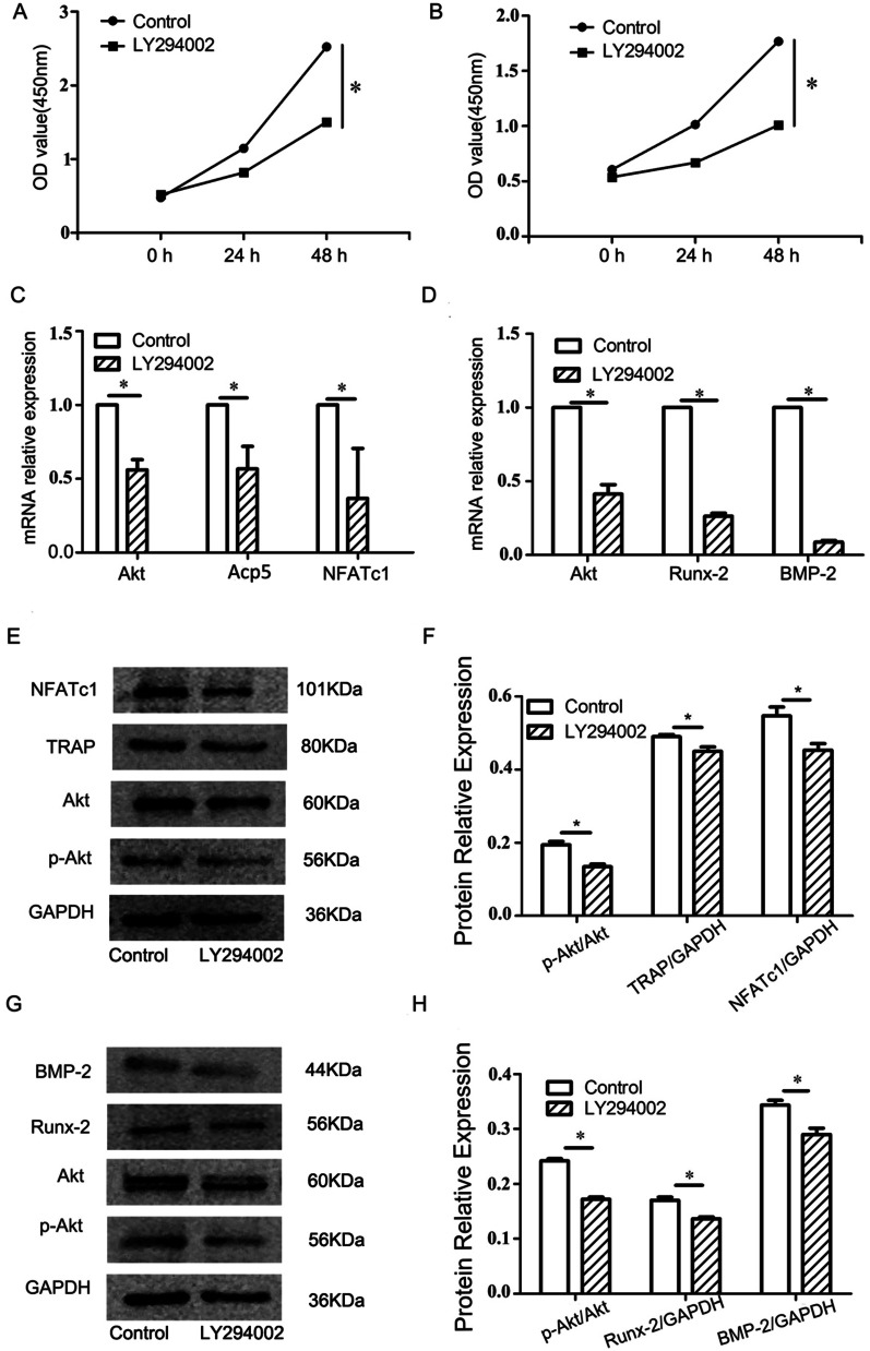
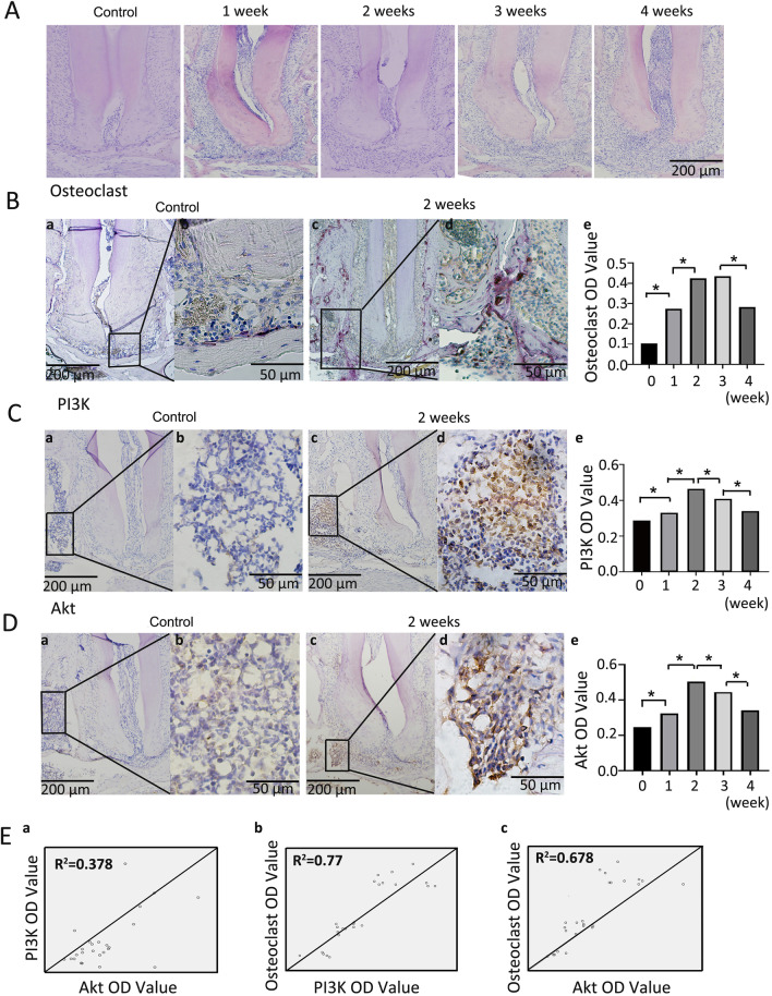
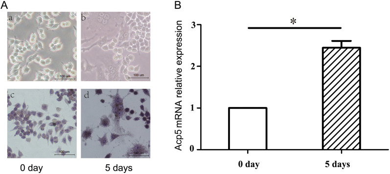
The relative mRNA expression of PI3K, Acp5 and NFATc1 in the normal human periodontal ligament and in chronic apical periodontitis were analyzed by real-time quantitative polymerase chain reaction (RT-qPCR). A mouse model of apical periodontitis was established by root canal exposure to the oral cavity, and HE staining was used to observe the progress of apical periodontitis. Immunohistochemical staining was used to detect the expression of PI3K and AKT in different stages of apical periodontitis, while enzymatic histochemical staining was used for detection of osteoclasts. An Escherichia coli lipopolysaccharide (LPS)-mediated inflammatory environment was also established at the osteoclast and osteoblast level, and osteoclasts or osteoblasts were treated with the PI3K inhibitor LY294002 to examine the role of PI3K in bone resorption.
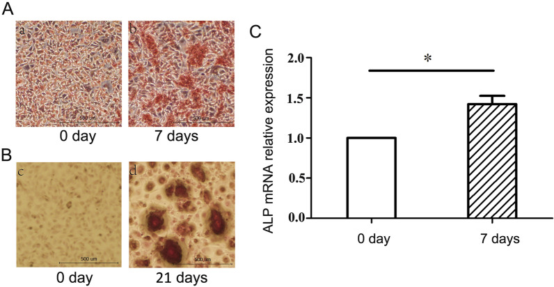
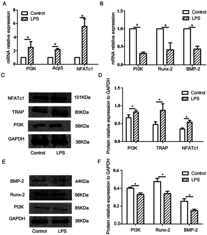
The expression of PI3K, Acp5 and NFATc1 genes in chronic apical periodontitis sample groups was significantly increased relative to healthy periodontal ligament tissue (P < 0.05). Mouse apical periodontitis was successfully established and bone resorption peaked between 2 and 3 weeks (P < 0.05). The expression of PI3K and Akt increased with the progression of inflammation, and reached a peak at 14 days (P < 0.05). The gene and protein expression of PI3K, TRAP and NFATc1 in osteoclasts were significantly increased (P < 0.05) in the E. coli LPS-mediated inflammatory microenvironment compared to the normal control group. Meanwhile in osteoblasts, the gene and protein expression of PI3K, BMP-2 and Runx2 were significantly reduced (P < 0.05) in the inflammatory microenvironment. With the addition of LY294002, expressions of bone resorption-related factors (TRAP, NFATc1) and bone formation-related factors (BMP-2, Runx2) significantly decreased (P < 0.05).
Under the inflammatory environment induced by LPS, PI3K participates in the occurrence and development of chronic apical periodontitis by regulating the proliferation and differentiation of osteoclasts and osteoblasts.
磷酸肌醇 3-激酶(PI3K)位于细胞内,参与调节细胞存活、增殖、凋亡和血管生成。本研究旨在探讨 PI3K 在根尖周炎骨破坏过程中的作用,为该病的治疗提供参考数据。
采用实时定量聚合酶链反应(RT-qPCR)分析正常牙周韧带和慢性根尖周炎中 PI3K、Acp5 和 NFATc1 的相对 mRNA 表达。通过根管暴露于口腔建立根尖周炎小鼠模型,采用 HE 染色观察根尖周炎的进展。采用免疫组织化学染色检测不同阶段根尖周炎中 PI3K 和 AKT 的表达,同时采用酶组织化学染色检测破骨细胞。还在破骨细胞和成骨细胞水平建立大肠杆菌脂多糖(LPS)介导的炎症环境,并使用 PI3K 抑制剂 LY294002 处理破骨细胞或成骨细胞,以研究 PI3K 在骨吸收中的作用。
与健康牙周韧带组织相比,慢性根尖周炎样本组中 PI3K、Acp5 和 NFATc1 基因的表达显著增加(P<0.05)。成功建立了小鼠根尖周炎,骨吸收在 2 至 3 周时达到峰值(P<0.05)。PI3K 和 Akt 的表达随着炎症的进展而增加,在 14 天时达到峰值(P<0.05)。在大肠杆菌 LPS 介导的炎症微环境中,破骨细胞中 PI3K、TRAP 和 NFATc1 的基因和蛋白表达明显增加(P<0.05),与正常对照组相比。同时,在成骨细胞中,炎症微环境中 PI3K、BMP-2 和 Runx2 的基因和蛋白表达明显降低(P<0.05)。加入 LY294002 后,骨吸收相关因子(TRAP、NFATc1)和骨形成相关因子(BMP-2、Runx2)的表达明显降低(P<0.05)。
在 LPS 诱导的炎症环境下,PI3K 通过调节破骨细胞和成骨细胞的增殖和分化参与慢性根尖周炎的发生和发展。