Xue Qiang, Wang Linbo, Zhao Yuanyu, Tong Wusong, Wang Jiancun, Li Gaoyi, Cheng Wei, Gao Liang, Dong Yan
Department of Neurosurgery, Eastern Hepatobiliary Surgery Hospital, Navy Medical University, Shanghai 200433, China.
Institute of Science and Technology for Brain-inspired Intelligence, Fudan University, Shanghai 210023, China.
J Clin Med. 2022 Jul 29;11(15):4421. doi: 10.3390/jcm11154421.
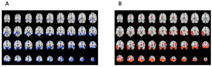
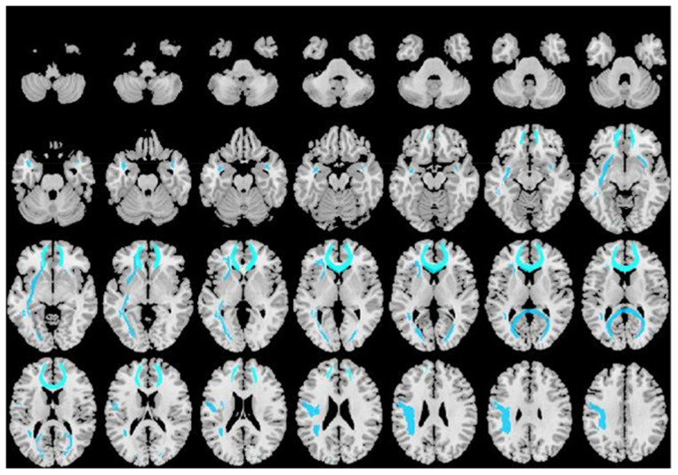
: Traumatic brain injury (TBI) often results in persistent cognitive impairment and psychiatric symptoms, while lesion location and severity are not consistent with its clinical complaints. Previous studies found cognitive deficits and psychiatric disorders following TBI are considered to be associated with prefrontal and medial temporal lobe lesions, however, the location and extent of contusions often cannot fully explain the patient's impairments. Thus, we try to find the structural changes of gray matter (GM) and white matter (WM), clarify their correlation with psychiatric symptoms and memory following TBI, and determine the brain regions that primary correlate with clinical measurements. : Overall, 32 TBI individuals and 23 healthy controls were recruited in the study. Cognitive impairment and psychiatric symptoms were examined by Mini-Mental State Examination (MMSE), Hospital Anxiety and Depression Scale (HADS), and Wechsler Memory Scale-Chinese Revision (WMS-CR). All MRI data were scanned using a Siemens Prisma 3.0 Tesla MRI system. T1 MRI data and diffusion tensor imaging (DTI) data were processed to analyze GM volume and WM microstructure separately. : In the present study, TBI patients underwent widespread decrease of GM volume in both cortical and subcortical regions. Among these regions, four brain areas including the left inferior temporal gyrus and medial temporal lobe, supplementary motor area, thalamus, and anterior cingulate cortex (ACC) were highly implicated in the post-traumatic cognitive impairment and psychiatric complaints. TBI patients also underwent changes of WM microstructure, involving decreased fractional anisotropy (FA) value in widespread WM tracts and increased mean diffusivity (MD) value in the forceps minor. The changes of WM microstructure were significantly correlated with the decrease of GM volume. : TBI causes widespread cortical and subcortical alterations including a reduction in GM volume and change in WM microstructure related to clinical manifestation. Lesions in temporal lobe may lead to more serious cognitive and emotional dysfunction, which should attract our high clinical attention.
创伤性脑损伤(TBI)常导致持续性认知障碍和精神症状,而损伤部位和严重程度与其临床症状并不一致。既往研究发现,TBI后的认知缺陷和精神障碍被认为与前额叶和内侧颞叶病变有关,然而,挫伤的部位和范围往往不能完全解释患者的损伤情况。因此,我们试图找出灰质(GM)和白质(WM)的结构变化,阐明它们与TBI后精神症状和记忆的相关性,并确定与临床测量主要相关的脑区。
总体而言,本研究招募了32名TBI患者和23名健康对照。通过简易精神状态检查表(MMSE)、医院焦虑抑郁量表(HADS)和韦氏记忆量表中文版(WMS-CR)对认知障碍和精神症状进行检查。所有MRI数据均使用西门子Prisma 3.0特斯拉MRI系统进行扫描。分别对T1 MRI数据和扩散张量成像(DTI)数据进行处理,以分析GM体积和WM微观结构。
在本研究中,TBI患者的皮质和皮质下区域GM体积普遍减少。在这些区域中,包括左侧颞下回和内侧颞叶、辅助运动区、丘脑和前扣带回皮质(ACC)在内的四个脑区与创伤后认知障碍和精神症状高度相关。TBI患者的WM微观结构也发生了变化,包括广泛WM束中的各向异性分数(FA)值降低,以及小钳夹中的平均扩散率(MD)值增加。WM微观结构的变化与GM体积的减少显著相关。
TBI会导致广泛的皮质和皮质下改变,包括GM体积减少和与临床表现相关的WM微观结构变化。颞叶病变可能导致更严重的认知和情感功能障碍,应引起我们高度的临床关注。