Thomas Lord Department of Computer Science, Viterbi School of Engineering, University of Southern California, Los Angeles, CA, 90089, USA.
Ethel Percy Andrus Gerontology Center, Leonard Davis School of Gerontology, University of Southern California, Los Angeles, CA, 90089, USA.
Neuroinformatics. 2024 Oct;22(4):591-606. doi: 10.1007/s12021-024-09694-2. Epub 2024 Nov 6.
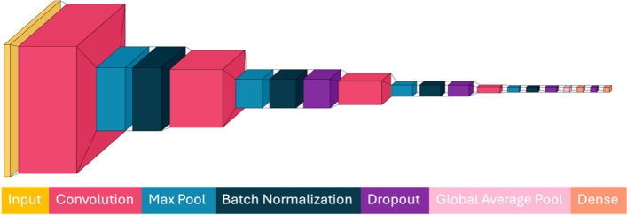
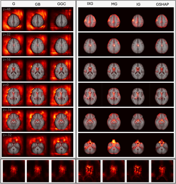
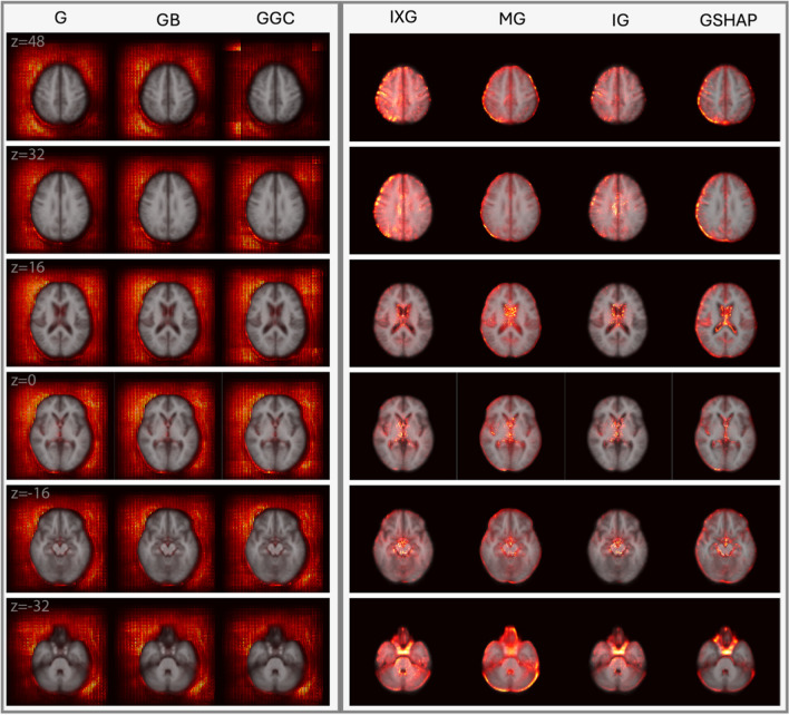
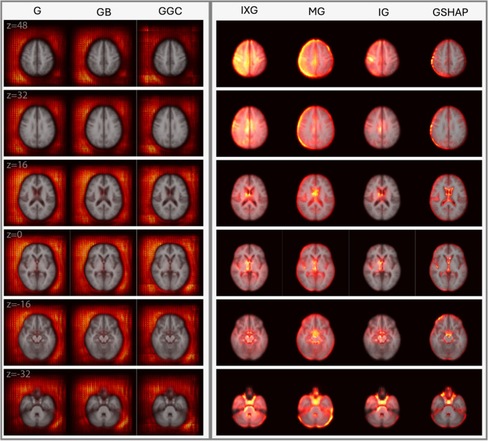
The black box nature of deep neural networks (DNNs) makes researchers and clinicians hesitant to rely on their findings. Saliency maps can enhance DNN explainability by suggesting the anatomic localization of relevant brain features. This study compares seven popular attribution-based saliency approaches to assign neuroanatomic interpretability to DNNs that estimate biological brain age (BA) from magnetic resonance imaging (MRI). Cognitively normal (CN) adults (N = 13,394, 5,900 males; mean age: 65.82 ± 8.89 years) are included for DNN training, testing, validation, and saliency map generation to estimate BA. To study saliency robustness to the presence of anatomic deviations from normality, saliency maps are also generated for adults with mild traumatic brain injury (mTBI, = 214, 135 males; mean age: 55.3 ± 9.9 years). We assess saliency methods' capacities to capture known anatomic features of brain aging and compare them to a surrogate ground truth whose anatomic saliency is known a priori. Anatomic aging features are identified most reliably by the integrated gradients method, which outperforms all others through its ability to localize relevant anatomic features. Gradient Shapley additive explanations, input × gradient, and masked gradient perform less consistently but still highlight ubiquitous neuroanatomic features of aging (ventricle dilation, hippocampal atrophy, sulcal widening). Saliency methods involving gradient saliency, guided backpropagation, and guided gradient-weight class attribution mapping localize saliency outside the brain, which is undesirable. Our research suggests the relative tradeoffs of saliency methods to interpret DNN findings during BA estimation in typical aging and after mTBI.
深度神经网络(DNN)的黑箱性质使得研究人员和临床医生对依赖其结果犹豫不决。显著图可以通过提示相关大脑特征的解剖定位来增强 DNN 的可解释性。本研究比较了七种流行的基于归因的显著方法,以便为从磁共振成像(MRI)估计生物脑龄(BA)的 DNN 赋予神经解剖学可解释性。认知正常(CN)成年人(N=13394,5900 名男性;平均年龄:65.82±8.89 岁)被纳入 DNN 训练、测试、验证和显著图生成,以估计 BA。为了研究显著图对存在解剖偏离正常的稳健性,还为轻度创伤性脑损伤(mTBI,=214,135 名男性;平均年龄:55.3±9.9 岁)的成年人生成显著图。我们评估了显著方法捕捉大脑老化已知解剖特征的能力,并将其与预先知道解剖显著度的替代真实值进行比较。集成梯度方法最可靠地识别出显著图的解剖老化特征,其通过定位相关解剖特征的能力优于所有其他方法。梯度 Shapley 加性解释、输入×梯度和掩蔽梯度的性能不太一致,但仍突出了老化的普遍神经解剖特征(脑室扩张、海马萎缩、脑沟增宽)。涉及梯度显著、引导反向传播和引导梯度权重类归因映射的显著方法将显著度定位在大脑之外,这是不理想的。我们的研究表明,在典型老化和 mTBI 后,在估计 BA 时,解释 DNN 结果的显著方法存在相对权衡。