• 文献检索
  • 文档翻译
  • 深度研究
  • 学术资讯
  • Suppr Zotero 插件Zotero 插件
  • 邀请有礼
  • 套餐&价格
  • 历史记录
应用&插件
Suppr Zotero 插件Zotero 插件浏览器插件Mac 客户端Windows 客户端微信小程序
定价
高级版会员购买积分包购买API积分包
服务
文献检索文档翻译深度研究API 文档MCP 服务
关于我们
关于 Suppr公司介绍联系我们用户协议隐私条款
关注我们

Suppr 超能文献

核心技术专利:CN118964589B侵权必究
粤ICP备2023148730 号-1Suppr @ 2026

文献检索

告别复杂PubMed语法,用中文像聊天一样搜索,搜遍4000万医学文献。AI智能推荐,让科研检索更轻松。

立即免费搜索

文件翻译

保留排版,准确专业,支持PDF/Word/PPT等文件格式,支持 12+语言互译。

免费翻译文档

深度研究

AI帮你快速写综述,25分钟生成高质量综述,智能提取关键信息,辅助科研写作。

立即免费体验

胎儿生长受限队列中的脑保护模式

Patterns of Brain Sparing in a Fetal Growth Restriction Cohort.

作者信息

Steller Jon G, Gumina Diane, Driver Camille, Peek Emma, Galan Henry L, Reeves Shane, Hobbins John C

机构信息

Division of Maternal Fetal Medicine, Department of Obstetrics and Gynecology, University of Colorado School of Medicine, Aurora, CO 80045, USA.

出版信息

J Clin Med. 2022 Aug 1;11(15):4480. doi: 10.3390/jcm11154480.

DOI:10.3390/jcm11154480
PMID:35956097
原文链接:https://pmc.ncbi.nlm.nih.gov/articles/PMC9369342/
Abstract

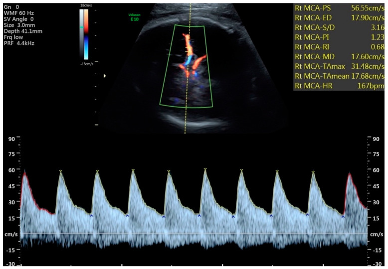
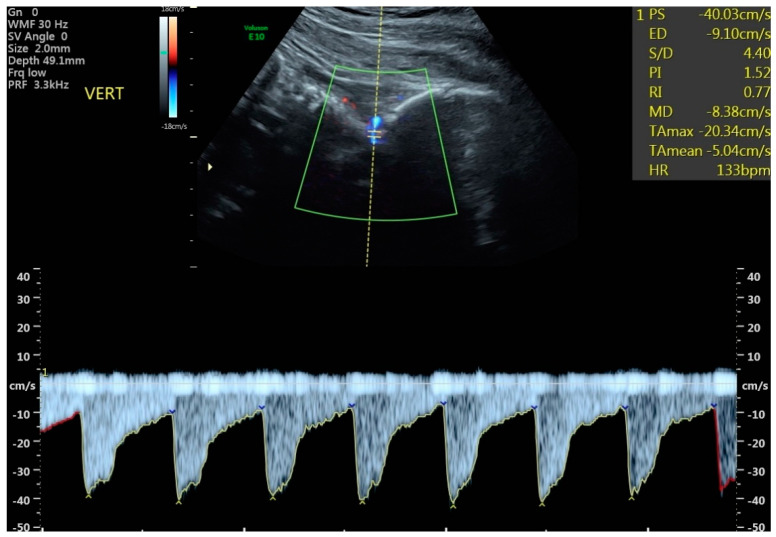
Objective: Our objective was to compare differences in Doppler blood flow in four fetal intracranial blood vessels in fetuses with late-onset fetal growth restriction (FGR) vs. those with small for gestational age (SGA). Methods: Fetuses with estimated fetal weight (EFW) <10th percentile were divided into SGA (n = 30) and FGR (n = 51) via Delphi criteria and had Doppler waveforms obtained from the middle cerebral artery (MCA), anterior cerebral artery (ACA), posterior cerebral artery (PCA), and vertebral artery (VA). A pulsatility index (PI) <5th centile was considered “abnormal”. Outcomes included birth metrics and neonatal intensive care unit (NICU) admission. Results: There were more abnormal cerebral vessel PIs in the FGR group versus the SGA group (36 vs. 4; p = 0.055). In FGR, ACA + MCA vessel abnormalities outnumbered PCA + VA abnormalities. All 8 fetuses with abnormal VA PIs had at least one other abnormal vessel. Fetuses with abnormal VA PIs had lower BW (1712 vs. 2500 g; p < 0.0001), delivered earlier (35.22 vs. 37.89 wks; p = 0.0052), and had more admissions to the NICU (71.43% vs. 24.44%; p = 0.023). Conclusions: There were more anterior vessels showing vasodilation than posterior vessels, but when the VA was abnormal, the fetuses were more severely affected clinically than those showing normal VA PIs.

摘要

目的

我们的目的是比较晚发性胎儿生长受限(FGR)胎儿与小于胎龄儿(SGA)胎儿颅内四条血管的多普勒血流差异。方法:根据德尔菲标准,将估计胎儿体重(EFW)<第10百分位数的胎儿分为SGA组(n = 30)和FGR组(n = 51),并获取大脑中动脉(MCA)、大脑前动脉(ACA)、大脑后动脉(PCA)和椎动脉(VA)的多普勒波形。搏动指数(PI)<第5百分位数被认为“异常”。结局指标包括出生指标和新生儿重症监护病房(NICU)入院情况。结果:FGR组的脑血管PI异常比SGA组更多(36例对4例;p = 0.055)。在FGR中,ACA + MCA血管异常多于PCA + VA血管异常。所有8例VA PI异常的胎儿至少还有一条其他血管异常。VA PI异常的胎儿出生体重较低(1712对2500 g;p < 0.0001),分娩更早(35.22对37.89周;p = 0.0052),且NICU入院率更高(71.43%对24.44%;p = 0.023)。结论:显示血管舒张的前循环血管比后循环血管更多,但当VA异常时,胎儿在临床上比VA PI正常的胎儿受影响更严重。

https://cdn.ncbi.nlm.nih.gov/pmc/blobs/8cf8/9369342/8842c7aa0a9d/jcm-11-04480-g002.jpg
https://cdn.ncbi.nlm.nih.gov/pmc/blobs/8cf8/9369342/6662a72d7912/jcm-11-04480-g001.jpg
https://cdn.ncbi.nlm.nih.gov/pmc/blobs/8cf8/9369342/8842c7aa0a9d/jcm-11-04480-g002.jpg
https://cdn.ncbi.nlm.nih.gov/pmc/blobs/8cf8/9369342/6662a72d7912/jcm-11-04480-g001.jpg
https://cdn.ncbi.nlm.nih.gov/pmc/blobs/8cf8/9369342/8842c7aa0a9d/jcm-11-04480-g002.jpg

相似文献

1
Patterns of Brain Sparing in a Fetal Growth Restriction Cohort.胎儿生长受限队列中的脑保护模式
J Clin Med. 2022 Aug 1;11(15):4480. doi: 10.3390/jcm11154480.
2
Ten-year experience of protocol-based management of small-for-gestational-age fetuses: perinatal outcome in late-pregnancy cases diagnosed after 32 weeks.基于方案的小胎龄儿管理十年经验:32 周后诊断的晚期妊娠病例的围生期结局。
Ultrasound Obstet Gynecol. 2021 Jan;57(1):62-69. doi: 10.1002/uog.23537.
3
Third-trimester growth diversity in small fetuses classified as appropriate-for-gestational age or small-for-gestational age at birth.出生时分类为适于胎龄或小于胎龄的小胎儿在孕晚期的生长差异。
Ultrasound Obstet Gynecol. 2021 Dec;58(6):882-891. doi: 10.1002/uog.23688.
4
Role of Doppler ultrasound at time of diagnosis of late-onset fetal growth restriction in predicting adverse perinatal outcome: prospective cohort study.多普勒超声在诊断晚发型胎儿生长受限中的作用对预测不良围生期结局的前瞻性队列研究。
Ultrasound Obstet Gynecol. 2020 Jun;55(6):793-798. doi: 10.1002/uog.20406. Epub 2020 May 8.
5
Fetal cardiac function in late-onset intrauterine growth restriction vs small-for-gestational age, as defined by estimated fetal weight, cerebroplacental ratio and uterine artery Doppler.根据估计胎儿体重、脑胎盘比率和子宫动脉多普勒所定义,晚发性宫内生长受限与小于胎龄儿的胎儿心脏功能
Ultrasound Obstet Gynecol. 2015 Oct;46(4):465-71. doi: 10.1002/uog.14930.
6
Fetal neurosonography detects differences in cortical development and corpus callosum in late-onset small fetuses.胎儿神经超声检查可检测出晚发型小胎儿皮质发育和胼胝体的差异。
Ultrasound Obstet Gynecol. 2021 Jul;58(1):42-47. doi: 10.1002/uog.23592.
7
Fetal growth restriction and intra-uterine growth restriction: guidelines for clinical practice from the French College of Gynaecologists and Obstetricians.胎儿生长受限与宫内生长受限:法国妇产科医师学院临床实践指南
Eur J Obstet Gynecol Reprod Biol. 2015 Oct;193:10-8. doi: 10.1016/j.ejogrb.2015.06.021. Epub 2015 Jul 2.
8
Assessment of ventricular contractility in fetuses with an estimated fetal weight less than the tenth centile.评估估计胎儿体重低于第十个百分位数的胎儿心室收缩力。
Am J Obstet Gynecol. 2019 Nov;221(5):498.e1-498.e22. doi: 10.1016/j.ajog.2019.05.042. Epub 2019 May 30.
9
Hemodynamic factors associated with fetal cardiac remodeling in late fetal growth restriction: a prospective study.与晚期胎儿生长受限胎儿心脏重构相关的血流动力学因素:一项前瞻性研究。
J Perinat Med. 2019 Sep 25;47(7):683-688. doi: 10.1515/jpm-2019-0217.
10
First-trimester and combined first- and second-trimester prediction of small-for-gestational age and late fetal growth restriction.早孕期及早中孕期联合预测小于胎龄儿及早中孕期晚期胎儿生长受限
Ultrasound Obstet Gynecol. 2019 Jan;53(1):55-61. doi: 10.1002/uog.19055. Epub 2018 Nov 26.

引用本文的文献

1
Fetal MRI study of brain differences in early-onset fetal growth restriction versus healthy controls at 30 weeks of gestation.孕30周时早发型胎儿生长受限与健康对照胎儿大脑差异的胎儿磁共振成像研究。
Eur J Obstet Gynecol Reprod Biol X. 2025 Jul 3;27:100417. doi: 10.1016/j.eurox.2025.100417. eCollection 2025 Sep.
2
How maternal position affects umbilical and middle cerebral artery Doppler indices: insights from a scoping review.母亲体位如何影响脐动脉和大脑中动脉多普勒指数:一项范围综述的见解
Rev Bras Ginecol Obstet. 2025 Jul 15;47. doi: 10.61622/rbgo/2025rbgo45. eCollection 2025.
3
Fetal brain development in fetal growth restriction using MRI: a systematic review.

本文引用的文献

1
ISUOG Practice Guidelines: diagnosis and management of small-for-gestational-age fetus and fetal growth restriction.国际妇产科超声学会(ISUOG)实践指南:小于胎龄儿和胎儿生长受限的诊断与管理
Ultrasound Obstet Gynecol. 2020 Aug;56(2):298-312. doi: 10.1002/uog.22134.
2
Society for Maternal-Fetal Medicine Consult Series #52: Diagnosis and management of fetal growth restriction: (Replaces Clinical Guideline Number 3, April 2012).母胎医学会咨询系列第 52 号:胎儿生长受限的诊断与处理:(替代 2012 年 4 月临床指南第 3 号)。
Am J Obstet Gynecol. 2020 Oct;223(4):B2-B17. doi: 10.1016/j.ajog.2020.05.010. Epub 2020 May 12.
3
使用磁共振成像(MRI)研究胎儿生长受限中的胎儿脑发育:一项系统综述。
BMC Pregnancy Childbirth. 2025 Feb 26;25(1):208. doi: 10.1186/s12884-024-07124-4.
4
Placental nanoparticle-mediated IGF1 gene therapy corrects fetal growth restriction in a guinea pig model.胎盘纳米颗粒介导的IGF1基因疗法可纠正豚鼠模型中的胎儿生长受限。
Gene Ther. 2024 Dec 4. doi: 10.1038/s41434-024-00508-3.
5
Diagnosis, Prevention, and Management of Fetal Growth Restriction (FGR).胎儿生长受限(FGR)的诊断、预防与管理
J Pers Med. 2024 Jun 28;14(7):698. doi: 10.3390/jpm14070698.
6
Does fetal growth restriction induce neuropathology within the developing brainstem?胎儿生长受限是否会引起发育中脑干的神经病理学改变?
J Physiol. 2023 Nov;601(21):4667-4689. doi: 10.1113/JP284191. Epub 2023 Aug 17.
7
Relationship between fetal middle cerebral artery pulsatility index and cerebroplacental ratio with adverse neonatal outcomes in low-risk pregnancy candidates for elective cesarean section: A cross-sectional study.择期剖宫产低风险妊娠孕妇胎儿大脑中动脉搏动指数与脑胎盘比率与不良新生儿结局的关系:一项横断面研究
Int J Reprod Biomed. 2022 Sep 6;20(8):663-670. doi: 10.18502/ijrm.v20i8.11755. eCollection 2022 Aug.
Size and shape of the four-chamber view of the fetal heart in fetuses with an estimated fetal weight less than the tenth centile.
胎儿心脏四腔心切面在估计胎儿体重小于第十个百分位的胎儿中的大小和形态。
Am J Obstet Gynecol. 2019 Nov;221(5):495.e1-495.e9. doi: 10.1016/j.ajog.2019.06.008. Epub 2019 Jun 14.
4
Assessment of ventricular contractility in fetuses with an estimated fetal weight less than the tenth centile.评估估计胎儿体重低于第十个百分位数的胎儿心室收缩力。
Am J Obstet Gynecol. 2019 Nov;221(5):498.e1-498.e22. doi: 10.1016/j.ajog.2019.05.042. Epub 2019 May 30.
5
ACOG Practice Bulletin No. 204: Fetal Growth Restriction.美国妇产科医师学会临床实践通告第 204 号:胎儿生长受限。
Obstet Gynecol. 2019 Feb;133(2):e97-e109. doi: 10.1097/AOG.0000000000003070.
6
Fetal Medicine Foundation fetal and neonatal population weight charts.胎儿医学基金会胎儿和新生儿人群体重图表。
Ultrasound Obstet Gynecol. 2018 Jul;52(1):44-51. doi: 10.1002/uog.19073. Epub 2018 May 30.
7
Long-term cardiovascular consequences of fetal growth restriction: biology, clinical implications, and opportunities for prevention of adult disease.胎儿生长受限的长期心血管后果:生物学、临床意义和预防成人疾病的机会。
Am J Obstet Gynecol. 2018 Feb;218(2S):S869-S879. doi: 10.1016/j.ajog.2017.12.012.
8
Fetal and umbilical Doppler ultrasound in high-risk pregnancies.高危妊娠中的胎儿及脐部多普勒超声检查
Cochrane Database Syst Rev. 2017 Jun 13;6(6):CD007529. doi: 10.1002/14651858.CD007529.pub4.
9
Committee Opinion No 700: Methods for Estimating the Due Date.委员会意见 700:预产期估计方法。
Obstet Gynecol. 2017 May;129(5):e150-e154. doi: 10.1097/AOG.0000000000002046.
10
Computing the Z Score and Centiles for Cross-sectional Analysis: A Practical Approach.计算用于横断面分析的Z分数和百分位数:一种实用方法。
J Ultrasound Med. 2017 Mar;36(3):459-473. doi: 10.7863/ultra.16.03025. Epub 2017 Jan 17.