• 文献检索
  • 文档翻译
  • 深度研究
  • 学术资讯
  • Suppr Zotero 插件Zotero 插件
  • 邀请有礼
  • 套餐&价格
  • 历史记录
应用&插件
Suppr Zotero 插件Zotero 插件浏览器插件Mac 客户端Windows 客户端微信小程序
定价
高级版会员购买积分包购买API积分包
服务
文献检索文档翻译深度研究API 文档MCP 服务
关于我们
关于 Suppr公司介绍联系我们用户协议隐私条款
关注我们

Suppr 超能文献

核心技术专利:CN118964589B侵权必究
粤ICP备2023148730 号-1Suppr @ 2026

文献检索

告别复杂PubMed语法,用中文像聊天一样搜索,搜遍4000万医学文献。AI智能推荐,让科研检索更轻松。

立即免费搜索

文件翻译

保留排版,准确专业,支持PDF/Word/PPT等文件格式,支持 12+语言互译。

免费翻译文档

深度研究

AI帮你快速写综述,25分钟生成高质量综述,智能提取关键信息,辅助科研写作。

立即免费体验

胸腔影像学创新:CT、放射组学、人工智能和 X 射线流速测定。

Innovations in thoracic imaging: CT, radiomics, AI and x-ray velocimetry.

机构信息

Department of Radiology, University of Groningen, University Medical Center Groningen, Groningen, the Netherlands.

Data Science in Health (DASH), University of Groningen, University Medical Center Groningen, Groningen, the Netherlands.

出版信息

Respirology. 2022 Oct;27(10):818-833. doi: 10.1111/resp.14344. Epub 2022 Aug 14.

DOI:10.1111/resp.14344
PMID:35965430
原文链接:https://pmc.ncbi.nlm.nih.gov/articles/PMC9546393/
Abstract

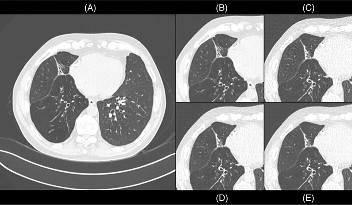
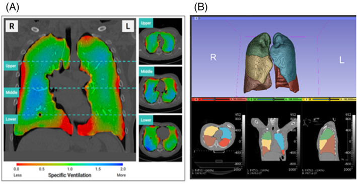
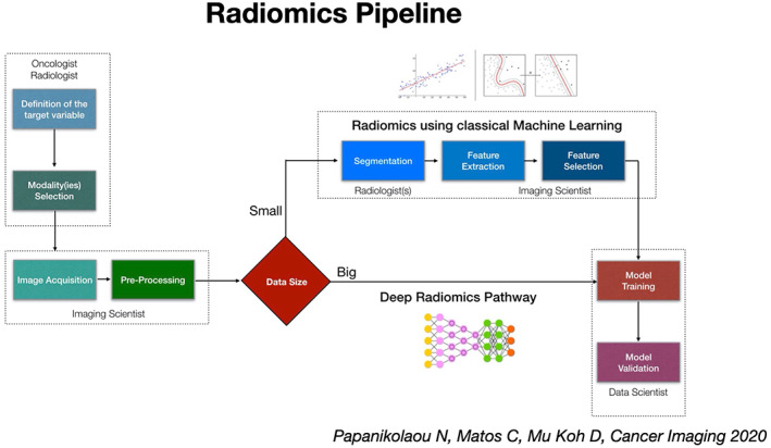
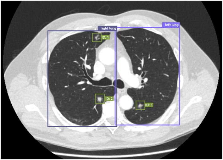
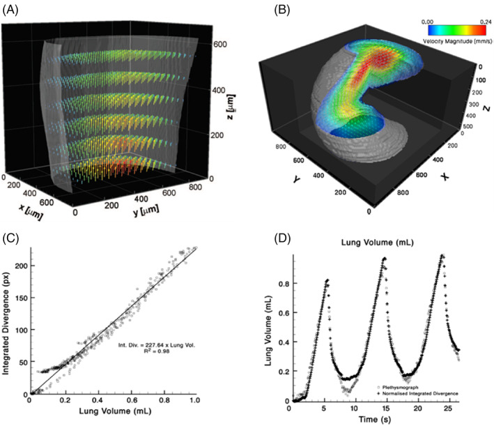
In recent years, pulmonary imaging has seen enormous progress, with the introduction, validation and implementation of new hardware and software. There is a general trend from mere visual evaluation of radiological images to quantification of abnormalities and biomarkers, and assessment of 'non visual' markers that contribute to establishing diagnosis or prognosis. Important catalysts to these developments in thoracic imaging include new indications (like computed tomography [CT] lung cancer screening) and the COVID-19 pandemic. This review focuses on developments in CT, radiomics, artificial intelligence (AI) and x-ray velocimetry for imaging of the lungs. Recent developments in CT include the potential for ultra-low-dose CT imaging for lung nodules, and the advent of a new generation of CT systems based on photon-counting detector technology. Radiomics has demonstrated potential towards predictive and prognostic tasks particularly in lung cancer, previously not achievable by visual inspection by radiologists, exploiting high dimensional patterns (mostly texture related) on medical imaging data. Deep learning technology has revolutionized the field of AI and as a result, performance of AI algorithms is approaching human performance for an increasing number of specific tasks. X-ray velocimetry integrates x-ray (fluoroscopic) imaging with unique image processing to produce quantitative four dimensional measurement of lung tissue motion, and accurate calculations of lung ventilation.

摘要

近年来,肺部影像学取得了巨大的进展,新的硬件和软件不断被引入、验证和应用。目前的趋势是从单纯的影像学图像的视觉评估,转变为对异常和生物标志物进行量化,以及评估有助于建立诊断或预后的“非视觉”标志物。推动这些胸部影像学发展的重要因素包括新的适应症(如 CT 肺癌筛查)和 COVID-19 大流行。本综述重点介绍了 CT、放射组学、人工智能 (AI) 和 X 射线流速测量在肺部成像中的进展。CT 的最新进展包括对肺结节进行超低剂量 CT 成像的可能性,以及基于光子计数探测器技术的新一代 CT 系统的问世。放射组学在预测和预后任务中表现出潜力,尤其是在肺癌方面,这是以前放射科医生仅凭视觉检查无法实现的,它利用了医学成像数据中的高维模式(主要与纹理有关)。深度学习技术彻底改变了人工智能领域,其结果是,人工智能算法在越来越多的特定任务中的性能正在接近人类水平。X 射线流速测量将 X 射线(透视)成像与独特的图像处理相结合,对肺组织运动进行定量的四维测量,并对肺通气进行精确计算。

https://cdn.ncbi.nlm.nih.gov/pmc/blobs/35a6/9546393/3dc908338892/RESP-27-818-g001.jpg
https://cdn.ncbi.nlm.nih.gov/pmc/blobs/35a6/9546393/0d78a58603d7/RESP-27-818-g004.jpg
https://cdn.ncbi.nlm.nih.gov/pmc/blobs/35a6/9546393/d3fe7b5f8abf/RESP-27-818-g005.jpg
https://cdn.ncbi.nlm.nih.gov/pmc/blobs/35a6/9546393/220e185edd43/RESP-27-818-g002.jpg
https://cdn.ncbi.nlm.nih.gov/pmc/blobs/35a6/9546393/223f6a7b2114/RESP-27-818-g003.jpg
https://cdn.ncbi.nlm.nih.gov/pmc/blobs/35a6/9546393/3dc908338892/RESP-27-818-g001.jpg

相似文献

1
Innovations in thoracic imaging: CT, radiomics, AI and x-ray velocimetry.胸腔影像学创新:CT、放射组学、人工智能和 X 射线流速测定。
Respirology. 2022 Oct;27(10):818-833. doi: 10.1111/resp.14344. Epub 2022 Aug 14.
2
Artificial intelligence-driven assessment of radiological images for COVID-19.人工智能驱动的 COVID-19 放射影像评估。
Comput Biol Med. 2021 Sep;136:104665. doi: 10.1016/j.compbiomed.2021.104665. Epub 2021 Jul 21.
3
Localized thin-section CT with radiomics feature extraction and machine learning to classify early-detected pulmonary nodules from lung cancer screening.基于影像组学特征提取和机器学习的局部薄层 CT 对肺癌筛查中早期检出肺结节的分类
Phys Med Biol. 2018 Mar 14;63(6):065005. doi: 10.1088/1361-6560/aaafab.
4
Artificial intelligence: A critical review of applications for lung nodule and lung cancer.人工智能:对肺结节和肺癌应用的批判性综述。
Diagn Interv Imaging. 2023 Jan;104(1):11-17. doi: 10.1016/j.diii.2022.11.007. Epub 2022 Dec 10.
5
A narrative review on characterization of acute respiratory distress syndrome in COVID-19-infected lungs using artificial intelligence.人工智能在 COVID-19 感染肺部中对急性呼吸窘迫综合征特征的叙述性综述。
Comput Biol Med. 2021 Mar;130:104210. doi: 10.1016/j.compbiomed.2021.104210. Epub 2021 Jan 18.
6
[Performance of Deep-learning-based Artificial Intelligence on Detection of Pulmonary Nodules in Chest CT].基于深度学习的人工智能在胸部CT肺结节检测中的性能
Zhongguo Fei Ai Za Zhi. 2019 Jun 20;22(6):336-340. doi: 10.3779/j.issn.1009-3419.2019.06.02.
7
[Artificial intelligence in lung imaging].[肺部成像中的人工智能]
Radiologe. 2020 Jan;60(1):42-47. doi: 10.1007/s00117-019-00611-2.
8
Artificial intelligence and radiomics enhance the positive predictive value of digital chest tomosynthesis for lung cancer detection within SOS clinical trial.人工智能和放射组学提高 SOS 临床试验中数字胸部断层合成术检测肺癌的阳性预测值。
Eur Radiol. 2020 Jul;30(7):4134-4140. doi: 10.1007/s00330-020-06783-z. Epub 2020 Mar 12.
9
[The contribution of artificial intelligence (AI) subsequent to the processing of thoracic imaging].[人工智能(AI)在胸部影像处理后的贡献]
Rev Mal Respir. 2024 Feb;41(2):110-126. doi: 10.1016/j.rmr.2023.12.001. Epub 2023 Dec 20.
10
Preoperative diagnosis of malignant pulmonary nodules in lung cancer screening with a radiomics nomogram.肺癌筛查中基于放射组学列线图的恶性肺结节术前诊断。
Cancer Commun (Lond). 2020 Jan;40(1):16-24. doi: 10.1002/cac2.12002. Epub 2020 Mar 3.

引用本文的文献

1
Contrast-Enhanced Cardiac Computed Tomography and the Presence of Intravascular Air: A Patient Safety Study.对比增强心脏计算机断层扫描与血管内空气的存在:一项患者安全研究。
J Clin Med. 2025 Jul 8;14(14):4842. doi: 10.3390/jcm14144842.
2
Future Applications of Cardiothoracic CT.心胸CT的未来应用
Radiology. 2025 Jun;315(3):e240085. doi: 10.1148/radiol.240085.
3
Application research of artificial intelligence software in the analysis of thyroid nodule ultrasound image characteristics.人工智能软件在甲状腺结节超声图像特征分析中的应用研究

本文引用的文献

1
Application of deep learning image reconstruction in low-dose chest CT scan.深度学习图像重建在低剂量胸部 CT 扫描中的应用。
Br J Radiol. 2022 May 1;95(1133):20210380. doi: 10.1259/bjr.20210380. Epub 2022 Jan 31.
2
Deep Learning Reconstruction Shows Better Lung Nodule Detection for Ultra-Low-Dose Chest CT.深度学习重建对超低剂量胸部 CT 显示出更好的肺结节检测效果。
Radiology. 2022 Apr;303(1):202-212. doi: 10.1148/radiol.210551. Epub 2022 Jan 18.
3
Pulmonary functional MRI: Detecting the structure-function pathologies that drive asthma symptoms and quality of life.
PLoS One. 2025 Jun 2;20(6):e0323343. doi: 10.1371/journal.pone.0323343. eCollection 2025.
4
A Thorough Review of the Clinical Applications of Artificial Intelligence in Lung Cancer.人工智能在肺癌临床应用的全面综述
Cancers (Basel). 2025 Mar 4;17(5):882. doi: 10.3390/cancers17050882.
5
Functional Lung Imaging Identifies Peripheral Ventilation Changes in ꞵ-ENaC Mice.功能性肺成像可识别β-ENaC小鼠外周通气变化。
Respirology. 2025 Apr;30(4):335-345. doi: 10.1111/resp.70009. Epub 2025 Feb 25.
6
Artificial Intelligence-Empowered Radiology-Current Status and Critical Review.人工智能赋能的放射学——现状与批判性综述
Diagnostics (Basel). 2025 Jan 24;15(3):282. doi: 10.3390/diagnostics15030282.
7
A low-dose CT reconstruction method using sub-pixel anisotropic diffusion.一种使用亚像素各向异性扩散的低剂量CT重建方法。
Nan Fang Yi Ke Da Xue Xue Bao. 2025 Jan 20;45(1):162-169. doi: 10.12122/j.issn.1673-4254.2025.01.19.
8
Deep learning-based image domain reconstruction enhances image quality and pulmonary nodule detection in ultralow-dose CT with adaptive statistical iterative reconstruction-V.基于深度学习的图像域重建可提高采用自适应统计迭代重建-V的超低剂量CT的图像质量及肺结节检测能力。
Eur Radiol. 2025 Jan 10. doi: 10.1007/s00330-024-11317-y.
9
Chest computed tomography for patients with sepsis in the emergency intensive care unit.急诊重症监护病房脓毒症患者的胸部计算机断层扫描。
Sci Data. 2024 Nov 20;11(1):1261. doi: 10.1038/s41597-024-04132-z.
10
Current understanding of the impact of United States military airborne hazards and burn pit exposures on respiratory health.目前对美国军事空降危害和燃烧坑暴露对呼吸健康影响的认识。
Part Fibre Toxicol. 2024 Oct 21;21(1):43. doi: 10.1186/s12989-024-00606-5.
肺部功能磁共振成像:检测导致哮喘症状和生活质量的结构-功能病理变化。
Respirology. 2022 Feb;27(2):114-133. doi: 10.1111/resp.14197. Epub 2022 Jan 10.
4
Spectral Photon-Counting CT Technology in Chest Imaging.胸部成像中的光谱光子计数CT技术
J Clin Med. 2021 Dec 9;10(24):5757. doi: 10.3390/jcm10245757.
5
Deep Learning for Lung Cancer Detection on Screening CT Scans: Results of a Large-Scale Public Competition and an Observer Study with 11 Radiologists.基于筛查CT扫描的肺癌深度学习检测:一项大规模公开竞赛结果及与11位放射科医生的观察者研究
Radiol Artif Intell. 2021 Oct 27;3(6):e210027. doi: 10.1148/ryai.2021210027. eCollection 2021 Nov.
6
Federated learning for predicting clinical outcomes in patients with COVID-19.基于联邦学习的 COVID-19 患者临床结局预测
Nat Med. 2021 Oct;27(10):1735-1743. doi: 10.1038/s41591-021-01506-3. Epub 2021 Sep 15.
7
Current and emerging artificial intelligence applications in chest imaging: a pediatric perspective.当前和新兴的人工智能在胸部成像中的应用:儿科视角。
Pediatr Radiol. 2022 Oct;52(11):2120-2130. doi: 10.1007/s00247-021-05146-0. Epub 2021 Sep 1.
8
Personalized Chest Computed Tomography: Minimum Diagnostic Radiation Dose Levels for the Detection of Fibrosis, Nodules, and Pneumonia.个性化胸部计算机断层扫描:检测纤维化、结节和肺炎的最低诊断辐射剂量水平。
Invest Radiol. 2022 Mar 1;57(3):148-156. doi: 10.1097/RLI.0000000000000822.
9
Impact of Morphotype on Image Quality and Diagnostic Performance of Ultra-Low-Dose Chest CT.形态类型对超低剂量胸部CT图像质量及诊断性能的影响
J Clin Med. 2021 Jul 26;10(15):3284. doi: 10.3390/jcm10153284.
10
First Performance Evaluation of an Artificial Intelligence-Based Computer-Aided Detection System for Pulmonary Nodule Evaluation in Dual-Source Photon-Counting Detector CT at Different Low-Dose Levels.基于人工智能的计算机辅助检测系统在不同低剂量水平的双源光子计数探测器CT中对肺结节评估的首次性能评估
Invest Radiol. 2022 Feb 1;57(2):108-114. doi: 10.1097/RLI.0000000000000814.