Li Xinling, Guo Fangfang, Zhou Zhen, Zhang Fandong, Wang Qin, Peng Zhijun, Su Datong, Fan Yaguang, Wang Ying
Department of Radiology, Tianjin Medical University General Hospital, Tianjin 300052, China.
Department of Radiology, the First Affiliated Hospital of XinXiang Medical College, Xinxiang 453100, China.
Zhongguo Fei Ai Za Zhi. 2019 Jun 20;22(6):336-340. doi: 10.3779/j.issn.1009-3419.2019.06.02.
The detection of pulmonary nodules is a key step to achieving the early diagnosis and therapy of lung cancer. Deep learning based Artificial intelligence (AI) presents as the state of the art in the area of nodule detection, however, a validation with clinical data is necessary for further application. Therefore, the aim of this study is to evaluate the performance of AI in the detection of malignant and non-calcified nodules in chest CT.
Two hundred chest computed tomography (CT) data were randomly selected from a self-built nodule database from Tianjin Medical University General Hospital. Both the pathology confirmed lung cancers and the nodules in the process of follow-up were included. All CTs were processed by AI and the results were compared with that of radiologists retrieved from the original medical reports. The ground truths were further determined by two experienced radiologists. The size and characteristics of the nodules were evaluated as well. The sensitivity and false positive rate were used to evaluate the effectiveness of AI and radiologists in detecting nodules. The McNemar test was used to determine whether there was a significant difference.
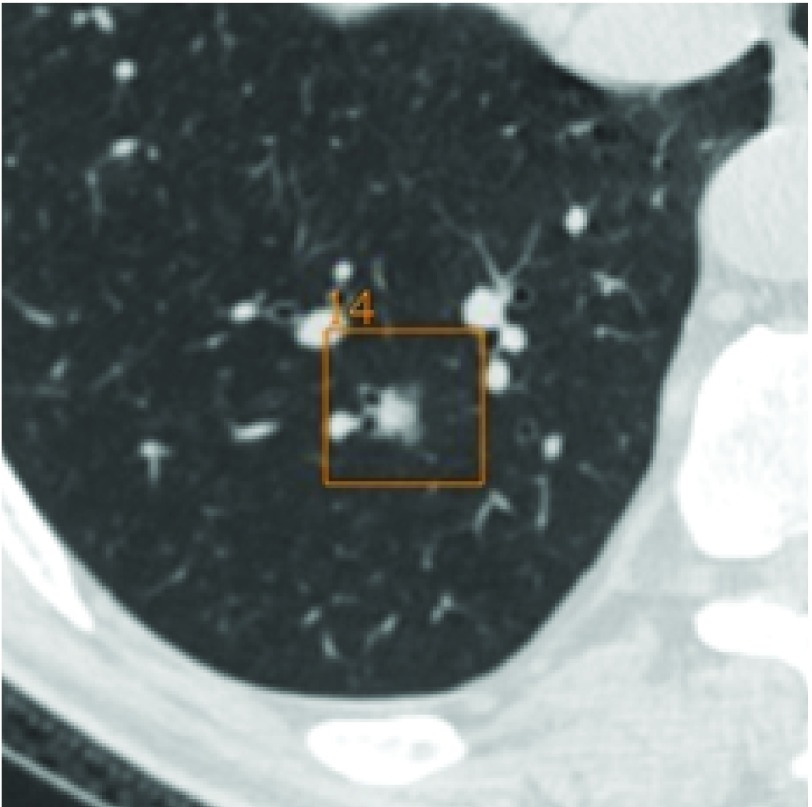
A total of 889 non-calcified nodules were determined by experts on chest CT, including 133 lung cancers. Of them, 442 nodules were less than 5 mm. The cancer detection rates of AI and radiologists are 100%. The sensitivity of AI on nodule detection was significantly higher than that of radiologists (99.1% vs 43%, P<0.001). The false-positive rate of AI was 4.9 per CT and decreased to 1.5 when nodules less than 5 mm were excluded.
AI achieves the detection of all malignancies and improve the sensitivity of pulmonary nodules detection beyond radiologists, with a low false positive rate after excluding small nodules.
肺结节的检测是实现肺癌早期诊断和治疗的关键步骤。基于深度学习的人工智能(AI)在结节检测领域代表了当前的先进水平,然而,要进一步应用还需要临床数据进行验证。因此,本研究的目的是评估AI在胸部CT中检测恶性和非钙化结节的性能。
从天津医科大学总医院自建的结节数据库中随机选取200例胸部计算机断层扫描(CT)数据。纳入病理确诊的肺癌以及随访过程中的结节。所有CT均由AI处理,并将结果与原始医学报告中的放射科医生的结果进行比较。真实情况由两名经验丰富的放射科医生进一步确定。还评估了结节的大小和特征。使用灵敏度和假阳性率来评估AI和放射科医生检测结节的有效性。采用McNemar检验来确定是否存在显著差异。
胸部CT专家共确定了889个非钙化结节,其中包括133例肺癌。其中,442个结节小于5mm。AI和放射科医生的癌症检测率均为100%。AI在结节检测方面的灵敏度显著高于放射科医生(99.1%对43%,P<0.001)。AI的假阳性率为每例CT 4.9个,排除小于5mm的结节后降至1.5个。
AI能够检测出所有恶性肿瘤,并且在检测肺结节方面比放射科医生提高了灵敏度,排除小结节后假阳性率较低。