• 文献检索
  • 文档翻译
  • 深度研究
  • 学术资讯
  • Suppr Zotero 插件Zotero 插件
  • 邀请有礼
  • 套餐&价格
  • 历史记录
应用&插件
Suppr Zotero 插件Zotero 插件浏览器插件Mac 客户端Windows 客户端微信小程序
定价
高级版会员购买积分包购买API积分包
服务
文献检索文档翻译深度研究API 文档MCP 服务
关于我们
关于 Suppr公司介绍联系我们用户协议隐私条款
关注我们

Suppr 超能文献

核心技术专利:CN118964589B侵权必究
粤ICP备2023148730 号-1Suppr @ 2026

文献检索

告别复杂PubMed语法,用中文像聊天一样搜索,搜遍4000万医学文献。AI智能推荐,让科研检索更轻松。

立即免费搜索

文件翻译

保留排版,准确专业,支持PDF/Word/PPT等文件格式,支持 12+语言互译。

免费翻译文档

深度研究

AI帮你快速写综述,25分钟生成高质量综述,智能提取关键信息,辅助科研写作。

立即免费体验

一种用于在临床环境中检测胸部X光片异常的人工智能系统的部署与验证。

Deployment and validation of an AI system for detecting abnormal chest radiographs in clinical settings.

作者信息

Nguyen Ngoc Huy, Nguyen Ha Quy, Nguyen Nghia Trung, Nguyen Thang Viet, Pham Hieu Huy, Nguyen Tuan Ngoc-Minh

机构信息

Phu Tho Department of Health, Viet Tri, Vietnam.

Medical Imaging Center, Vingroup Big Data Institute, Hanoi, Vietnam.

出版信息

Front Digit Health. 2022 Jul 27;4:890759. doi: 10.3389/fdgth.2022.890759. eCollection 2022.

DOI:10.3389/fdgth.2022.890759
PMID:35966141
原文链接:https://pmc.ncbi.nlm.nih.gov/articles/PMC9367219/
Abstract

BACKGROUND

The purpose of this paper is to demonstrate a mechanism for deploying and validating an AI-based system for detecting abnormalities on chest X-ray scans at the Phu Tho General Hospital, Vietnam. We aim to investigate the performance of the system in real-world clinical settings and compare its effectiveness to the in-lab performance.

METHOD

The AI system was directly integrated into the Hospital's Picture Archiving and Communication System (PACS) after being trained on a fixed annotated dataset from other sources. The system's performance was prospectively measured by matching and comparing the AI results with the radiology reports of 6,285 chest X-ray examinations extracted from the Hospital Information System (HIS) over the last 2 months of 2020. The normal/abnormal status of a radiology report was determined by a set of rules and served as the ground truth.

RESULTS

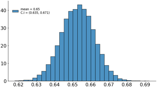
Our system achieves an F1 score-the harmonic average of the recall and the precision-of 0.653 (95% CI 0.635, 0.671) for detecting any abnormalities on chest X-rays. This corresponds to an accuracy of 79.6%, a sensitivity of 68.6%, and a specificity of 83.9%.

CONCLUSIONS

Computer-Aided Diagnosis (CAD) systems for chest radiographs using artificial intelligence (AI) have recently shown great potential as a second opinion for radiologists. However, the performances of such systems were mostly evaluated on a fixed dataset in a retrospective manner and, thus, far from the real performances in clinical practice. Despite a significant drop from the in-lab performance, our result establishes a reasonable level of confidence in applying such a system in real-life situations.

摘要

背景

本文旨在展示在越南富寿综合医院部署和验证基于人工智能的胸部X光扫描异常检测系统的机制。我们旨在研究该系统在实际临床环境中的性能,并将其有效性与实验室性能进行比较。

方法

在使用来自其他来源的固定注释数据集进行训练后,该人工智能系统被直接集成到医院的图像存档与通信系统(PACS)中。通过将人工智能结果与从医院信息系统(HIS)中提取的2020年最后两个月的6285份胸部X光检查的放射学报告进行匹配和比较,前瞻性地测量了该系统的性能。放射学报告的正常/异常状态由一组规则确定,并作为基本事实。

结果

我们的系统在检测胸部X光片上的任何异常时,F1分数(召回率和精确率的调和平均值)达到0.653(95%置信区间0.635,0.671)。这对应于79.6%的准确率、68.6%的灵敏度和83.9%的特异性。

结论

使用人工智能(AI)的胸部X光计算机辅助诊断(CAD)系统最近作为放射科医生的第二意见显示出巨大潜力。然而,此类系统的性能大多以回顾性方式在固定数据集上进行评估,因此与临床实践中的实际性能相差甚远。尽管与实验室性能相比有显著下降,但我们的结果为在实际情况中应用此类系统建立了合理的信心水平。

https://cdn.ncbi.nlm.nih.gov/pmc/blobs/f1fe/9367219/10ebcfab9fe0/fdgth-04-890759-g0006.jpg
https://cdn.ncbi.nlm.nih.gov/pmc/blobs/f1fe/9367219/5e7335645334/fdgth-04-890759-g0001.jpg
https://cdn.ncbi.nlm.nih.gov/pmc/blobs/f1fe/9367219/328f5db3fda6/fdgth-04-890759-g0002.jpg
https://cdn.ncbi.nlm.nih.gov/pmc/blobs/f1fe/9367219/df8567c871f2/fdgth-04-890759-g0003.jpg
https://cdn.ncbi.nlm.nih.gov/pmc/blobs/f1fe/9367219/695c4be4b090/fdgth-04-890759-g0004.jpg
https://cdn.ncbi.nlm.nih.gov/pmc/blobs/f1fe/9367219/899a044a29bc/fdgth-04-890759-g0005.jpg
https://cdn.ncbi.nlm.nih.gov/pmc/blobs/f1fe/9367219/10ebcfab9fe0/fdgth-04-890759-g0006.jpg

相似文献

1
Deployment and validation of an AI system for detecting abnormal chest radiographs in clinical settings.一种用于在临床环境中检测胸部X光片异常的人工智能系统的部署与验证。
Front Digit Health. 2022 Jul 27;4:890759. doi: 10.3389/fdgth.2022.890759. eCollection 2022.
2
Comparison of Chest Radiograph Interpretations by Artificial Intelligence Algorithm vs Radiology Residents.人工智能算法与放射科住院医师对胸部 X 线片解读的比较。
JAMA Netw Open. 2020 Oct 1;3(10):e2022779. doi: 10.1001/jamanetworkopen.2020.22779.
3
AI-based computer-aided diagnostic system of chest digital tomography synthesis: Demonstrating comparative advantage with X-ray-based AI systems.基于人工智能的胸部数字断层合成计算机辅助诊断系统:与基于 X 射线的人工智能系统比较优势展示。
Comput Methods Programs Biomed. 2023 Oct;240:107643. doi: 10.1016/j.cmpb.2023.107643. Epub 2023 Jun 5.
4
Development and validation of open-source deep neural networks for comprehensive chest x-ray reading: a retrospective, multicentre study.开源深度学习神经网络在全面胸部 X 线阅读中的开发和验证:一项回顾性、多中心研究。
Lancet Digit Health. 2024 Jan;6(1):e44-e57. doi: 10.1016/S2589-7500(23)00218-2. Epub 2023 Dec 8.
5
Role of an Automated Deep Learning Algorithm for Reliable Screening of Abnormality in Chest Radiographs: A Prospective Multicenter Quality Improvement Study.自动深度学习算法在胸部X光片异常可靠筛查中的作用:一项前瞻性多中心质量改进研究。
Diagnostics (Basel). 2022 Nov 7;12(11):2724. doi: 10.3390/diagnostics12112724.
6
Performance of an AI based CAD system in solid lung nodule detection on chest phantom radiographs compared to radiology residents and fellow radiologists.基于人工智能的计算机辅助检测(CAD)系统在胸部体模X光片上检测实性肺结节的性能与放射科住院医师和放射科专科医师的比较。
J Thorac Dis. 2021 May;13(5):2728-2737. doi: 10.21037/jtd-20-3522.
7
Artificial Intelligence-assisted chest X-ray assessment scheme for COVID-19.人工智能辅助 COVID-19 胸片评估方案。
Eur Radiol. 2021 Aug;31(8):6039-6048. doi: 10.1007/s00330-020-07628-5. Epub 2021 Jan 20.
8
Association of Artificial Intelligence-Aided Chest Radiograph Interpretation With Reader Performance and Efficiency.人工智能辅助的胸部 X 光片解读与读者表现和效率的关联。
JAMA Netw Open. 2022 Aug 1;5(8):e2229289. doi: 10.1001/jamanetworkopen.2022.29289.
9
Independent evaluation of the accuracy of 5 artificial intelligence software for detecting lung nodules on chest X-rays.对5种用于在胸部X光片上检测肺结节的人工智能软件准确性的独立评估。
Quant Imaging Med Surg. 2024 Aug 1;14(8):5288-5303. doi: 10.21037/qims-24-160. Epub 2024 Jul 25.
10
Artificial intelligence-supported lung cancer detection by multi-institutional readers with multi-vendor chest radiographs: a retrospective clinical validation study.人工智能支持的多机构阅片者对多厂家胸部 X 线片的肺癌检测:一项回顾性临床验证研究。
BMC Cancer. 2021 Oct 18;21(1):1120. doi: 10.1186/s12885-021-08847-9.

引用本文的文献

1
Clinical validation of an artificial intelligence algorithm for classifying tuberculosis and pulmonary findings in chest radiographs.用于胸部X光片中肺结核及肺部表现分类的人工智能算法的临床验证
Front Artif Intell. 2025 Feb 6;8:1512910. doi: 10.3389/frai.2025.1512910. eCollection 2025.
2
Deep Learning-Based Object Detection Strategies for Disease Detection and Localization in Chest X-Ray Images.基于深度学习的胸部X光图像疾病检测与定位目标检测策略
Diagnostics (Basel). 2024 Nov 22;14(23):2636. doi: 10.3390/diagnostics14232636.
3
Position Statements of the Emerging Trends Committee of the Asian Oceanian Society of Radiology on the Adoption and Implementation of Artificial Intelligence for Radiology.

本文引用的文献

1
A review of deep learning in medical imaging: Imaging traits, technology trends, case studies with progress highlights, and future promises.医学成像中的深度学习综述:成像特征、技术趋势、具有进展亮点的案例研究及未来展望。
Proc IEEE Inst Electr Electron Eng. 2021 May;109(5):820-838. doi: 10.1109/JPROC.2021.3054390. Epub 2021 Feb 26.
2
Causality-Inspired Single-Source Domain Generalization for Medical Image Segmentation.用于医学图像分割的因果启发式单源域泛化
IEEE Trans Med Imaging. 2023 Apr;42(4):1095-1106. doi: 10.1109/TMI.2022.3224067. Epub 2023 Apr 3.
3
VinDr-CXR: An open dataset of chest X-rays with radiologist's annotations.
亚洲大洋洲放射学会新兴趋势委员会关于采用和实施人工智能在放射学中的立场声明。
Korean J Radiol. 2024 Jul;25(7):603-612. doi: 10.3348/kjr.2024.0419.
4
Evaluation of the Performance of an Artificial Intelligence (AI) Algorithm in Detecting Thoracic Pathologies on Chest Radiographs.评估一种人工智能(AI)算法在胸部X光片上检测胸部病变的性能。
Diagnostics (Basel). 2024 Jun 4;14(11):1183. doi: 10.3390/diagnostics14111183.
5
A deep-learning-based framework for identifying and localizing multiple abnormalities and assessing cardiomegaly in chest X-ray.基于深度学习的框架,用于识别和定位胸部 X 光片中的多种异常情况并评估心胸比。
Nat Commun. 2024 Feb 14;15(1):1347. doi: 10.1038/s41467-024-45599-z.
6
Improving diagnosis accuracy with an intelligent image retrieval system for lung pathologies detection: a features extractor approach.利用智能图像检索系统提高肺部病变检测的诊断准确率:特征提取方法。
Sci Rep. 2023 Oct 3;13(1):16619. doi: 10.1038/s41598-023-42366-w.
7
Validation of a Deep Learning Model for Detecting Chest Pathologies from Digital Chest Radiographs.用于从数字化胸部X光片中检测胸部病变的深度学习模型的验证
Diagnostics (Basel). 2023 Feb 2;13(3):557. doi: 10.3390/diagnostics13030557.
8
Performance of a Chest Radiography AI Algorithm for Detection of Missed or Mislabeled Findings: A Multicenter Study.用于检测漏诊或错误标记结果的胸部X线人工智能算法的性能:一项多中心研究。
Diagnostics (Basel). 2022 Aug 28;12(9):2086. doi: 10.3390/diagnostics12092086.
VinDr-CXR:一个带有放射科医生标注的胸部 X 光数据集。
Sci Data. 2022 Jul 20;9(1):429. doi: 10.1038/s41597-022-01498-w.
4
Effect of a comprehensive deep-learning model on the accuracy of chest x-ray interpretation by radiologists: a retrospective, multireader multicase study.深度学习模型对放射科医师解读胸部 X 光片准确性的影响:一项回顾性、多读者多病例研究。
Lancet Digit Health. 2021 Aug;3(8):e496-e506. doi: 10.1016/S2589-7500(21)00106-0. Epub 2021 Jul 1.
5
Automated abnormality classification of chest radiographs using deep convolutional neural networks.使用深度卷积神经网络对胸部X光片进行自动异常分类。
NPJ Digit Med. 2020 May 14;3:70. doi: 10.1038/s41746-020-0273-z. eCollection 2020.
6
MIMIC-CXR, a de-identified publicly available database of chest radiographs with free-text reports.MIMIC-CXR,一个去标识化的、公开可用的、包含自由文本报告的胸部 X 光数据库。
Sci Data. 2019 Dec 12;6(1):317. doi: 10.1038/s41597-019-0322-0.
7
Chest Radiograph Interpretation with Deep Learning Models: Assessment with Radiologist-adjudicated Reference Standards and Population-adjusted Evaluation.深度学习模型在胸部 X 线片解读中的应用:使用经过放射科医师裁定的参考标准和人群校正评估进行评估。
Radiology. 2020 Feb;294(2):421-431. doi: 10.1148/radiol.2019191293. Epub 2019 Dec 3.
8
Human-machine partnership with artificial intelligence for chest radiograph diagnosis.人机协作利用人工智能进行胸部X光诊断。
NPJ Digit Med. 2019 Nov 18;2:111. doi: 10.1038/s41746-019-0189-7. eCollection 2019.
9
Efficient Deep Network Architectures for Fast Chest X-Ray Tuberculosis Screening and Visualization.高效的深度网络架构,用于快速的胸部 X 射线结核病筛查和可视化。
Sci Rep. 2019 Apr 18;9(1):6268. doi: 10.1038/s41598-019-42557-4.
10
Development and Validation of a Deep Learning-Based Automated Detection Algorithm for Major Thoracic Diseases on Chest Radiographs.基于深度学习的胸部 X 线片主要胸部疾病自动检测算法的开发与验证。
JAMA Netw Open. 2019 Mar 1;2(3):e191095. doi: 10.1001/jamanetworkopen.2019.1095.