• 文献检索
  • 文档翻译
  • 深度研究
  • 学术资讯
  • Suppr Zotero 插件Zotero 插件
  • 邀请有礼
  • 套餐&价格
  • 历史记录
应用&插件
Suppr Zotero 插件Zotero 插件浏览器插件Mac 客户端Windows 客户端微信小程序
定价
高级版会员购买积分包购买API积分包
服务
文献检索文档翻译深度研究API 文档MCP 服务
关于我们
关于 Suppr公司介绍联系我们用户协议隐私条款
关注我们

Suppr 超能文献

核心技术专利:CN118964589B侵权必究
粤ICP备2023148730 号-1Suppr @ 2026

文献检索

告别复杂PubMed语法,用中文像聊天一样搜索,搜遍4000万医学文献。AI智能推荐,让科研检索更轻松。

立即免费搜索

文件翻译

保留排版,准确专业,支持PDF/Word/PPT等文件格式,支持 12+语言互译。

免费翻译文档

深度研究

AI帮你快速写综述,25分钟生成高质量综述,智能提取关键信息,辅助科研写作。

立即免费体验

使用深度学习从4DCT心脏血管造影的容积再现中检测左心室壁运动异常。

Detection of left ventricular wall motion abnormalities from volume rendering of 4DCT cardiac angiograms using deep learning.

作者信息

Chen Zhennong, Contijoch Francisco, Colvert Gabrielle M, Manohar Ashish, Kahn Andrew M, Narayan Hari K, McVeigh Elliot

机构信息

Department of Bioengineering, UC San Diego School of Engineering, La Jolla, CA, United States.

Department of Radiology, UC San Diego School of Medicine, La Jolla, CA, United States.

出版信息

Front Cardiovasc Med. 2022 Jul 28;9:919751. doi: 10.3389/fcvm.2022.919751. eCollection 2022.

DOI:10.3389/fcvm.2022.919751
PMID:35966529
原文链接:https://pmc.ncbi.nlm.nih.gov/articles/PMC9366190/
Abstract

BACKGROUND

The presence of left ventricular (LV) wall motion abnormalities (WMA) is an independent indicator of adverse cardiovascular events in patients with cardiovascular diseases. We develop and evaluate the ability to detect cardiac wall motion abnormalities (WMA) from dynamic volume renderings (VR) of clinical 4D computed tomography (CT) angiograms using a deep learning (DL) framework.

METHODS

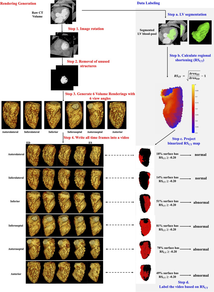
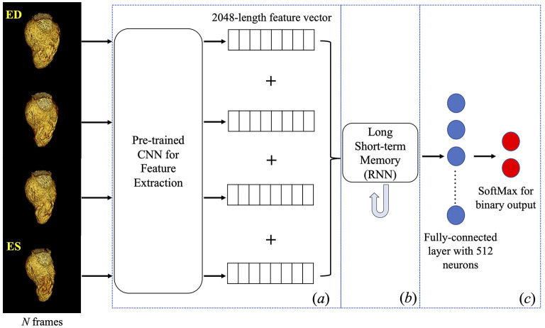
Three hundred forty-three ECG-gated cardiac 4DCT studies (age: 61 ± 15, 60.1% male) were retrospectively evaluated. Volume-rendering videos of the LV blood pool were generated from 6 different perspectives (i.e., six views corresponding to every 60-degree rotation around the LV long axis); resulting in 2058 unique videos. Ground-truth WMA classification for each video was performed by evaluating the extent of impaired regional shortening visible (measured in the original 4DCT data). DL classification of each video for the presence of WMA was performed by first extracting image features frame-by-frame using a pre-trained Inception network and then evaluating the set of features using a long short-term memory network. Data were split into 60% for 5-fold cross-validation and 40% for testing.

RESULTS

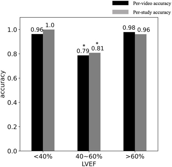
Volume rendering videos represent ~800-fold data compression of the 4DCT volumes. Per-video DL classification performance was high for both cross-validation (accuracy = 93.1%, sensitivity = 90.0% and specificity = 95.1%, κ: 0.86) and testing (90.9, 90.2, and 91.4% respectively, κ: 0.81). Per-study performance was also high (cross-validation: 93.7, 93.5, 93.8%, κ: 0.87; testing: 93.5, 91.9, 94.7%, κ: 0.87). By re-binning per-video results into the 6 regional views of the LV we showed DL was accurate (mean accuracy = 93.1 and 90.9% for cross-validation and testing cohort, respectively) for every region. DL classification strongly agreed (accuracy = 91.0%, κ: 0.81) with expert visual assessment.

CONCLUSIONS

Dynamic volume rendering of the LV blood pool combined with DL classification can accurately detect regional WMA from cardiac CT.

摘要

背景

左心室(LV)壁运动异常(WMA)的存在是心血管疾病患者发生不良心血管事件的独立指标。我们开发并评估了使用深度学习(DL)框架从临床四维计算机断层扫描(CT)血管造影的动态容积渲染(VR)中检测心脏壁运动异常(WMA)的能力。

方法

回顾性评估了343项心电图门控心脏4DCT研究(年龄:61±15岁,男性占60.1%)。从6个不同视角生成左心室血池的容积渲染视频(即围绕左心室长轴每旋转60度对应的六个视图);共产生2058个独特视频。通过评估原始4DCT数据中可见的局部缩短受损程度,对每个视频进行WMA分类的真实情况判定。通过首先使用预训练的Inception网络逐帧提取图像特征,然后使用长短期记忆网络评估特征集,对每个视频进行WMA存在情况的DL分类。数据分为60%用于5折交叉验证,40%用于测试。

结果

容积渲染视频代表4DCT容积约800倍的数据压缩。每个视频的DL分类性能在交叉验证(准确率=93.1%,灵敏度=90.0%,特异性=95.1%,κ值:0.86)和测试(分别为90.9%、90.2%和91.4%,κ值:0.81)中都很高。每项研究的性能也很高(交叉验证:93.7%、93.5%、93.8%,κ值:0.87;测试:93.5%、91.9%、94.7%,κ值:0.87)。通过将每个视频的结果重新分类到左心室的6个区域视图中,我们表明DL对每个区域都很准确(交叉验证队列和测试队列的平均准确率分别为93.1%和90.9%)。DL分类与专家视觉评估高度一致(准确率=91.0%,κ值:0.81)。

结论

左心室血池的动态容积渲染结合DL分类可以从心脏CT中准确检测局部WMA。

https://cdn.ncbi.nlm.nih.gov/pmc/blobs/422e/9366190/4c7dd10a35a0/fcvm-09-919751-g0003.jpg
https://cdn.ncbi.nlm.nih.gov/pmc/blobs/422e/9366190/a4affe5f541f/fcvm-09-919751-g0001.jpg
https://cdn.ncbi.nlm.nih.gov/pmc/blobs/422e/9366190/59e4b1cb0afb/fcvm-09-919751-g0002.jpg
https://cdn.ncbi.nlm.nih.gov/pmc/blobs/422e/9366190/4c7dd10a35a0/fcvm-09-919751-g0003.jpg
https://cdn.ncbi.nlm.nih.gov/pmc/blobs/422e/9366190/a4affe5f541f/fcvm-09-919751-g0001.jpg
https://cdn.ncbi.nlm.nih.gov/pmc/blobs/422e/9366190/59e4b1cb0afb/fcvm-09-919751-g0002.jpg
https://cdn.ncbi.nlm.nih.gov/pmc/blobs/422e/9366190/4c7dd10a35a0/fcvm-09-919751-g0003.jpg

相似文献

1
Detection of left ventricular wall motion abnormalities from volume rendering of 4DCT cardiac angiograms using deep learning.使用深度学习从4DCT心脏血管造影的容积再现中检测左心室壁运动异常。
Front Cardiovasc Med. 2022 Jul 28;9:919751. doi: 10.3389/fcvm.2022.919751. eCollection 2022.
2
Deep learning automates detection of wall motion abnormalities measurement of longitudinal strain from ECG-gated CT images.深度学习可自动检测壁运动异常并从心电图门控CT图像测量纵向应变。
Front Cardiovasc Med. 2022 Dec 15;9:1009445. doi: 10.3389/fcvm.2022.1009445. eCollection 2022.
3
Myocardial Regional Shortening from 4D Cardiac CT Angiography for the Detection of Left Ventricular Segmental Wall Motion Abnormality.用于检测左心室节段性壁运动异常的4D心脏CT血管造影术的心肌区域缩短
Radiol Cardiothorac Imaging. 2023 Mar 9;5(2):e220134. doi: 10.1148/ryct.220134. eCollection 2023 Apr.
4
Automated cardiac volume assessment and cardiac long- and short-axis imaging plane prediction from electrocardiogram-gated computed tomography volumes enabled by deep learning.通过深度学习实现的心电图门控计算机断层扫描容积自动心脏容积评估及心脏长轴和短轴成像平面预测。
Eur Heart J Digit Health. 2021 Mar 22;2(2):311-322. doi: 10.1093/ehjdh/ztab033. eCollection 2021 Jun.
5
High-Throughput Precision Phenotyping of Left Ventricular Hypertrophy With Cardiovascular Deep Learning.高通量精准表型分析左心室肥厚的心血管深度学习方法。
JAMA Cardiol. 2022 Apr 1;7(4):386-395. doi: 10.1001/jamacardio.2021.6059.
6
Deep learning-based left ventricular segmentation demonstrates improved performance on respiratory motion-resolved whole-heart reconstructions.基于深度学习的左心室分割在呼吸运动分辨全心脏重建上表现出了更好的性能。
Front Radiol. 2023 Jun 2;3:1144004. doi: 10.3389/fradi.2023.1144004. eCollection 2023.
7
Rapid assessment of left ventricular segmental wall motion, ejection fraction, and volumes with single breath-hold, multi-slice TrueFISP MR imaging.采用单屏气多层TrueFISP磁共振成像快速评估左心室节段性室壁运动、射血分数和容积。
J Cardiovasc Magn Reson. 2006;8(3):435-44. doi: 10.1080/10976640600575205.
8
Impact of persistent left ventricular regional wall motion abnormalities in childhood cancer survivors after anthracycline therapy: Assessment of global left ventricular myocardial performance by 3D speckle-tracking echocardiography.蒽环类药物治疗后儿童癌症幸存者持续性左心室局部壁运动异常的影响:通过三维斑点追踪超声心动图评估左心室整体心肌功能
J Cardiol. 2017 Oct;70(4):396-401. doi: 10.1016/j.jjcc.2016.12.015. Epub 2017 Feb 24.
9
Lung tumor segmentation in 4D CT images using motion convolutional neural networks.使用运动卷积神经网络进行 4D CT 图像中的肺部肿瘤分割。
Med Phys. 2021 Nov;48(11):7141-7153. doi: 10.1002/mp.15204. Epub 2021 Sep 13.
10
[Assessment of left ventricular function by gated cardiac blood-pool emission computed tomography using a rotating gamma camera].[使用旋转γ相机的门控心血池发射计算机断层扫描评估左心室功能]
Kaku Igaku. 1991 Jan;28(1):51-61.

引用本文的文献

1
Myocardial Regional Shortening from 4D Cardiac CT Angiography for the Detection of Left Ventricular Segmental Wall Motion Abnormality.用于检测左心室节段性壁运动异常的4D心脏CT血管造影术的心肌区域缩短
Radiol Cardiothorac Imaging. 2023 Mar 9;5(2):e220134. doi: 10.1148/ryct.220134. eCollection 2023 Apr.
2
Deep learning automates detection of wall motion abnormalities measurement of longitudinal strain from ECG-gated CT images.深度学习可自动检测壁运动异常并从心电图门控CT图像测量纵向应变。
Front Cardiovasc Med. 2022 Dec 15;9:1009445. doi: 10.3389/fcvm.2022.1009445. eCollection 2022.

本文引用的文献

1
Octree Representation Improves Data Fidelity of Cardiac CT Images and Convolutional Neural Network Semantic Segmentation of Left Atrial and Ventricular Chambers.八叉树表示法提高了心脏CT图像的数据保真度以及左心房和心室腔的卷积神经网络语义分割效果。
Radiol Artif Intell. 2021 Sep 29;3(6):e210036. doi: 10.1148/ryai.2021210036. eCollection 2021 Nov.
2
Novel 4DCT Method to Measure Regional Left Ventricular Endocardial Shortening Before and After Transcatheter Mitral Valve Implantation.测量经导管二尖瓣植入术前和术后局部左心室心内膜缩短的新型4DCT方法
Struct Heart. 2021;5(4):410-419. doi: 10.1080/24748706.2021.1934617. Epub 2021 Jul 15.
3
Automated cardiac volume assessment and cardiac long- and short-axis imaging plane prediction from electrocardiogram-gated computed tomography volumes enabled by deep learning.
通过深度学习实现的心电图门控计算机断层扫描容积自动心脏容积评估及心脏长轴和短轴成像平面预测。
Eur Heart J Digit Health. 2021 Mar 22;2(2):311-322. doi: 10.1093/ehjdh/ztab033. eCollection 2021 Jun.
4
Anthropomorphic left ventricular mesh phantom: a framework to investigate the accuracy of SQUEEZ using Coherent Point Drift for the detection of regional wall motion abnormalities.拟人化左心室网格模型:一种用于研究使用相干点漂移的SQUEEZ检测局部室壁运动异常准确性的框架。
J Med Imaging (Bellingham). 2019 Oct;6(4):045001. doi: 10.1117/1.JMI.6.4.045001. Epub 2019 Dec 10.
5
Identification and Quantification of Cardiovascular Structures From CCTA: An End-to-End, Rapid, Pixel-Wise, Deep-Learning Method.从 CCTA 中识别和量化心血管结构:一种端到端、快速、逐像素、深度学习方法。
JACC Cardiovasc Imaging. 2020 May;13(5):1163-1171. doi: 10.1016/j.jcmg.2019.08.025. Epub 2019 Oct 11.
6
Precise measurement of coronary stenosis diameter with CCTA using CT number calibration.利用 CT 数校准对冠状动脉狭窄程度进行精确的 CCTA 测量。
Med Phys. 2019 Dec;46(12):5514-5527. doi: 10.1002/mp.13862. Epub 2019 Oct 31.
7
Impact of apical foreshortening on deformation measurements: a report from the EACVI-ASE Strain Standardization Task Force.心尖部缩短对形变测量的影响:欧洲心血管影像学会-美国超声心动图学会应变标准化工作组的报告
Eur Heart J Cardiovasc Imaging. 2020 Mar 1;21(3):337-343. doi: 10.1093/ehjci/jez189.
8
Deep Learning for Diagnosis of Chronic Myocardial Infarction on Nonenhanced Cardiac Cine MRI.深度学习在非增强型心脏电影磁共振成像上诊断慢性心肌梗死的应用。
Radiology. 2019 Jun;291(3):606-617. doi: 10.1148/radiol.2019182304. Epub 2019 Apr 30.
9
Convolutional Neural Network Based Metal Artifact Reduction in X-Ray Computed Tomography.基于卷积神经网络的 X 射线计算机断层扫描中的金属伪影减少。
IEEE Trans Med Imaging. 2018 Jun;37(6):1370-1381. doi: 10.1109/TMI.2018.2823083.
10
Regional myocardial strain measurements from 4DCT in patients with normal LV function.四维 CT 测量正常左心室功能患者的局部心肌应变。
J Cardiovasc Comput Tomogr. 2018 Sep-Oct;12(5):372-378. doi: 10.1016/j.jcct.2018.05.002. Epub 2018 May 9.