• 文献检索
  • 文档翻译
  • 深度研究
  • 学术资讯
  • Suppr Zotero 插件Zotero 插件
  • 邀请有礼
  • 套餐&价格
  • 历史记录
应用&插件
Suppr Zotero 插件Zotero 插件浏览器插件Mac 客户端Windows 客户端微信小程序
定价
高级版会员购买积分包购买API积分包
服务
文献检索文档翻译深度研究API 文档MCP 服务
关于我们
关于 Suppr公司介绍联系我们用户协议隐私条款
关注我们

Suppr 超能文献

核心技术专利:CN118964589B侵权必究
粤ICP备2023148730 号-1Suppr @ 2026

文献检索

告别复杂PubMed语法,用中文像聊天一样搜索,搜遍4000万医学文献。AI智能推荐,让科研检索更轻松。

立即免费搜索

文件翻译

保留排版,准确专业,支持PDF/Word/PPT等文件格式,支持 12+语言互译。

免费翻译文档

深度研究

AI帮你快速写综述,25分钟生成高质量综述,智能提取关键信息,辅助科研写作。

立即免费体验

将绵羊大脑映射到人类大脑:绵羊脑图谱的必要性。

Mapping sheep to human brain: The need for a sheep brain atlas.

作者信息

Banstola Ashik, Reynolds John N J

机构信息

Department of Anatomy, School of Biomedical Sciences, University of Otago, Dunedin, New Zealand.

Brain Health Research Centre, University of Otago, Dunedin, New Zealand.

出版信息

Front Vet Sci. 2022 Jul 29;9:961413. doi: 10.3389/fvets.2022.961413. eCollection 2022.

DOI:10.3389/fvets.2022.961413
PMID:35967997
原文链接:https://pmc.ncbi.nlm.nih.gov/articles/PMC9372442/
Abstract

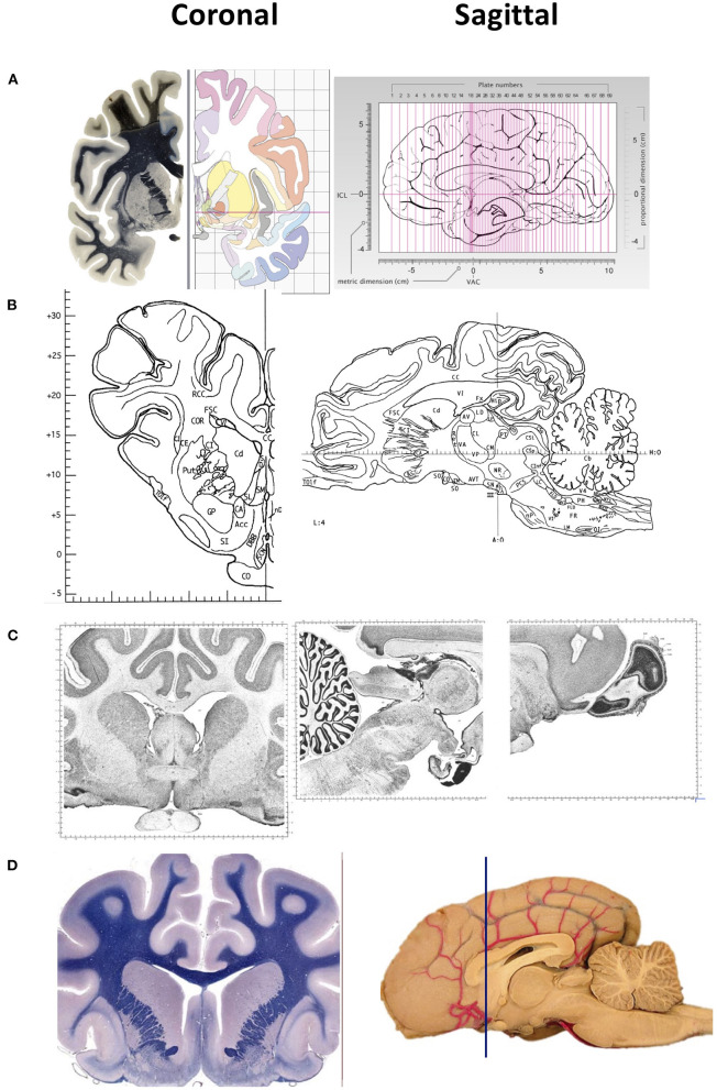
A brain atlas is essential for understanding the anatomical relationship between neuroanatomical structures. Standard stereotaxic coordinates and reference systems have been developed for humans, non-human primates and small laboratory animals to contribute to translational neuroscience research. Despite similar neuroanatomical and neurofunctional features between the sheep and human brain, little is known of the sheep brain stereotaxy, and a detailed sheep atlas is scarce. Here, we briefly discuss the value of using sheep in neurological research and the paucity of literature concerning the coordinates system during neurosurgical approaches. Recent advancements such as computerized tomography, positron emission tomography, magnetic resonance imaging, functional magnetic resonance imaging and diffusion tensor imaging are used for targeting and localizing the coordinates and brain areas in humans. Still, their application in sheep is rare due to the lack of a 3D stereotaxic sheep atlas by which to map sheep brain structures to its human counterparts. More recently, a T1- and T2-weighted high-resolution MRI 3D stereotaxic atlas of the sheep brain has been generated, however, the journey to create a sheep brain atlas by which to map directly to the human brain is still uncharted. Therefore, developing a detailed sheep brain atlas is valuable for the future to facilitate the use of sheep as a large animal experimental non-primate model for translational neurological research.

摘要

脑图谱对于理解神经解剖结构之间的解剖学关系至关重要。已为人类、非人灵长类动物和小型实验动物开发了标准立体定向坐标和参考系统,以促进转化神经科学研究。尽管绵羊和人类大脑在神经解剖学和神经功能特征上相似,但对绵羊脑立体定位知之甚少,详细的绵羊图谱也很稀少。在此,我们简要讨论在神经学研究中使用绵羊的价值以及神经外科手术方法中有关坐标系统的文献匮乏情况。诸如计算机断层扫描、正电子发射断层扫描、磁共振成像、功能磁共振成像和扩散张量成像等最新进展被用于在人类中定位坐标和脑区。然而,由于缺乏用于将绵羊脑结构映射到人类对应结构的三维立体定向绵羊图谱,它们在绵羊中的应用很少。最近,已经生成了绵羊脑的T1加权和T2加权高分辨率MRI三维立体定向图谱,但是创建一个可直接映射到人类大脑的绵羊脑图谱的征程仍未开启。因此,开发详细的绵羊脑图谱对于未来将绵羊用作转化神经学研究的大型动物实验非灵长类模型具有重要价值。

https://cdn.ncbi.nlm.nih.gov/pmc/blobs/9a2e/9372442/e2bf4904e438/fvets-09-961413-g0001.jpg

相似文献

1
Mapping sheep to human brain: The need for a sheep brain atlas.将绵羊大脑映射到人类大脑:绵羊脑图谱的必要性。
Front Vet Sci. 2022 Jul 29;9:961413. doi: 10.3389/fvets.2022.961413. eCollection 2022.
2
Computation of a high-resolution MRI 3D stereotaxic atlas of the sheep brain.绵羊脑高分辨率磁共振成像3D立体定向图谱的计算。
J Comp Neurol. 2017 Feb 15;525(3):676-692. doi: 10.1002/cne.24079. Epub 2016 Aug 9.
3
A stereotaxic breed-averaged, symmetric T2w canine brain atlas including detailed morphological and volumetrical data sets.一种基于立体定向的、具有品种平均性的、对称的 T2w 犬脑图谱,包括详细的形态学和容积数据集。
Neuroimage. 2019 Feb 15;187:93-103. doi: 10.1016/j.neuroimage.2018.01.066. Epub 2018 Jan 31.
4
A stereotaxic, population-averaged T1w ovine brain atlas including cerebral morphology and tissue volumes.一个包含脑形态和组织体积的立体定向、群体平均T1加权绵羊脑图谱。
Front Neuroanat. 2015 Jun 4;9:69. doi: 10.3389/fnana.2015.00069. eCollection 2015.
5
Diffusion Tensor Magnetic Resonance Tractography of the Sheep Brain: An Atlas of the Ovine White Matter Fiber Bundles.绵羊脑的扩散张量磁共振纤维束成像:绵羊白质纤维束图谱
Front Vet Sci. 2019 Oct 16;6:345. doi: 10.3389/fvets.2019.00345. eCollection 2019.
6
A stereotaxic MRI template set for the rat brain with tissue class distribution maps and co-registered anatomical atlas: application to pharmacological MRI.一套用于大鼠脑的立体定向磁共振成像模板集,带有组织类别分布图和共同配准的解剖图谱:在药物磁共振成像中的应用。
Neuroimage. 2006 Aug 15;32(2):538-50. doi: 10.1016/j.neuroimage.2006.04.214. Epub 2006 Jun 19.
7
A stereotaxic template atlas of the macaque brain for digital imaging and quantitative neuroanatomy.用于数字成像和定量神经解剖学的猕猴脑立体定位模板图谱。
Neuroimage. 1996 Oct;4(2):119-50. doi: 10.1006/nimg.1996.0036.
8
Stereotactic localization of the human pedunculopontine nucleus: atlas-based coordinates and validation of a magnetic resonance imaging protocol for direct localization.人类脚桥核的立体定向定位:基于图谱的坐标及直接定位磁共振成像方案的验证
Brain. 2008 Jun;131(Pt 6):1588-98. doi: 10.1093/brain/awn075. Epub 2008 May 8.
9
An MRI based average macaque monkey stereotaxic atlas and space (MNI monkey space).基于 MRI 的恒河猴平均脑立体定位图谱及空间(MNI 猴空间)。
Neuroimage. 2011 Apr 15;55(4):1435-42. doi: 10.1016/j.neuroimage.2011.01.040. Epub 2011 Jan 20.
10
Stereotaxic F-FDG PET and MRI templates with three-dimensional digital atlas for statistical parametric mapping analysis of tree shrew brain.树鼩脑的立体定位 F-FDG PET 和 MRI 模板与三维数字图谱用于统计参数映射分析。
J Neurosci Methods. 2018 Jan 1;293:105-116. doi: 10.1016/j.jneumeth.2017.09.006. Epub 2017 Sep 14.

引用本文的文献

1
Absolute depth-resolved optic axis measurement with catheter-based polarization sensitive optical coherence tomography.基于导管的偏振敏感光学相干断层扫描进行绝对深度分辨光轴测量。
Biomed Opt Express. 2024 Nov 25;15(12):6957-6976. doi: 10.1364/BOE.538560. eCollection 2024 Dec 1.
2
Long-term hypoxia modulates depolarization activation of BK currents in fetal sheep middle cerebral arterial myocytes.长期缺氧调节胎羊大脑中动脉肌细胞中BK电流的去极化激活。
Front Physiol. 2024 Nov 5;15:1479882. doi: 10.3389/fphys.2024.1479882. eCollection 2024.
3
The prototypical interferonopathy: Aicardi-Goutières syndrome from bedside to bench.

本文引用的文献

1
The orbitofrontal cortex of the sheep. Topography, organization, neurochemistry, digital tensor imaging and comparison with the chimpanzee and human.绵羊的眶额皮质。地形学、组织学、神经化学、数字张量成像及与黑猩猩和人类的比较。
Brain Struct Funct. 2022 Jun;227(5):1871-1891. doi: 10.1007/s00429-022-02479-w. Epub 2022 Mar 26.
2
The primary visual cortex of Cetartiodactyls: organization, cytoarchitectonics and comparison with perissodactyls and primates.偶蹄目动物的初级视皮层:组织、细胞构筑学及与奇蹄目动物和灵长类动物的比较。
Brain Struct Funct. 2022 May;227(4):1195-1225. doi: 10.1007/s00429-021-02392-8. Epub 2021 Oct 3.
3
典型的干扰素病:从床边到实验室的 Aicardi-Goutières 综合征。
Immunol Rev. 2024 Oct;327(1):83-99. doi: 10.1111/imr.13413. Epub 2024 Oct 29.
4
Application and Safety of Externally Controlled Metronomic Drug Delivery to the Brain by an Implantable Smart Pump in a Sheep Model.在绵羊模型中通过植入式智能泵对大脑进行外部控制的节律性药物递送的应用与安全性
Neurosurgery. 2025 Feb 1;96(2):471-478. doi: 10.1227/neu.0000000000003155. Epub 2024 Sep 6.
5
Beyond the surface: how diffusion-weighted imaging reveals large animal brain microstructure and connectivity.透过表象:扩散加权成像如何揭示大型动物大脑微观结构和连通性
Front Neurosci. 2024 Jun 26;18:1411982. doi: 10.3389/fnins.2024.1411982. eCollection 2024.
6
Sheep (Ovis aries) training protocol for voluntary awake and unrestrained structural brain MRI acquisitions.绵羊(Ovis aries)用于自愿清醒和不受约束的结构脑 MRI 采集的训练方案。
Behav Res Methods. 2024 Oct;56(7):7761-7773. doi: 10.3758/s13428-024-02449-6. Epub 2024 Jun 21.
7
FAST (Flexible Acetylcholine Sensing Thread): Real-Time Detection of Acetylcholine with a Flexible Solid-Contact Potentiometric Sensor.FAST(柔性乙酰胆碱传感线):利用柔性固体接触电位传感器实时检测乙酰胆碱
Bioengineering (Basel). 2023 May 27;10(6):655. doi: 10.3390/bioengineering10060655.
8
Anatomy of hypothalamic and diencephalic nuclei involved in seasonal fertility regulation in ewes.参与母羊季节性繁殖调控的下丘脑和间脑核团的解剖结构。
Front Vet Sci. 2023 Feb 16;10:1101024. doi: 10.3389/fvets.2023.1101024. eCollection 2023.
9
Harnessing Ultrasound for Targeting Drug Delivery to the Brain and Breaching the Blood-Brain Tumour Barrier.利用超声将药物靶向递送至大脑并突破血脑肿瘤屏障。
Pharmaceutics. 2022 Oct 19;14(10):2231. doi: 10.3390/pharmaceutics14102231.
10
The Sheep as a Large Animal Model for the Investigation and Treatment of Human Disorders.绵羊作为用于人类疾病研究与治疗的大型动物模型。
Biology (Basel). 2022 Aug 23;11(9):1251. doi: 10.3390/biology11091251.
Stereotactic Cortical Atlas of the Domestic Canine Brain.
犬脑立体定向皮质图谱
Sci Rep. 2020 Mar 16;10(1):4781. doi: 10.1038/s41598-020-61665-0.
4
The development of brain magnetic resonance approaches in large animal models for preclinical research.用于临床前研究的大型动物模型中脑磁共振方法的发展。
Anim Front. 2019 Jun 25;9(3):44-51. doi: 10.1093/af/vfz024. eCollection 2019 Jul.
5
Diffusion Tensor Magnetic Resonance Tractography of the Sheep Brain: An Atlas of the Ovine White Matter Fiber Bundles.绵羊脑的扩散张量磁共振纤维束成像:绵羊白质纤维束图谱
Front Vet Sci. 2019 Oct 16;6:345. doi: 10.3389/fvets.2019.00345. eCollection 2019.
6
The motor cortex of the sheep: laminar organization, projections and diffusion tensor imaging of the intracranial pyramidal and extrapyramidal tracts.绵羊的运动皮层:颅内锥体束和锥体外束的层状组织、投射和扩散张量成像。
Brain Struct Funct. 2019 Jun;224(5):1933-1946. doi: 10.1007/s00429-019-01885-x. Epub 2019 May 14.
7
Chemical neuroanatomy of the substantia nigra in the ovine brain.绵羊脑内黑质的化学神经解剖学。
J Chem Neuroanat. 2019 Apr;97:43-56. doi: 10.1016/j.jchemneu.2019.01.007. Epub 2019 Jan 18.
8
Loss of interneurons and disruption of perineuronal nets in the cerebral cortex following hypoxia-ischaemia in near-term fetal sheep.近足月胎羊缺氧缺血后脑皮质中间神经元丢失和周围神经网破坏。
Sci Rep. 2018 Dec 6;8(1):17686. doi: 10.1038/s41598-018-36083-y.
9
Computed tomography provides enhanced techniques for longitudinal monitoring of progressive intracranial volume loss associated with regional neurodegeneration in ovine neuronal ceroid lipofuscinoses.计算机断层扫描为羊神经元蜡样脂褐质沉积症中与区域性神经退行性变相关的进行性颅内容积损失的纵向监测提供了增强技术。
Brain Behav. 2018 Sep;8(9):e01096. doi: 10.1002/brb3.1096. Epub 2018 Aug 23.
10
Establishing sheep as an experimental species to validate ultrasound-mediated blood-brain barrier opening for potential therapeutic interventions.建立绵羊作为实验动物模型,验证超声介导的血脑屏障开放用于潜在的治疗干预。
Theranostics. 2018 Apr 3;8(9):2583-2602. doi: 10.7150/thno.22852. eCollection 2018.