Molecular Pathology, School of Dentistry Universidad de la República (UDELAR) Las Heras 1925, Montevideo 14600, Uruguay
Med Oral Patol Oral Cir Bucal. 2022 Sep 1;27(5):e403-e409. doi: 10.4317/medoral.25145.
Ep-CAM, a transmembrane glycoprotein expressed in most epithelium in normal conditions, has diverse roles in these tissues, including in cell adhesion, proliferation, differentiation, cell cycle regulation, migration and intracellular signaling. It is also over-expressed in most malignant neoplasia, participating in the initiation, progression, and metastatic dissemination of the tumor. The expression and roles of this protein in oral neoplasia, particularly in odontogenic tumors, remain unestablished. The objective of this study consisted in analyzing the expression of this protein in ameloblastoma and tooth germ.
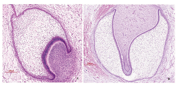
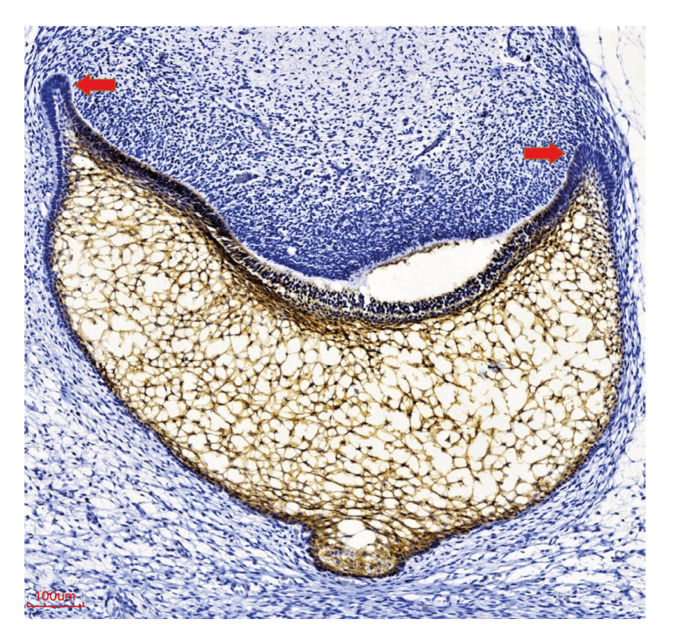
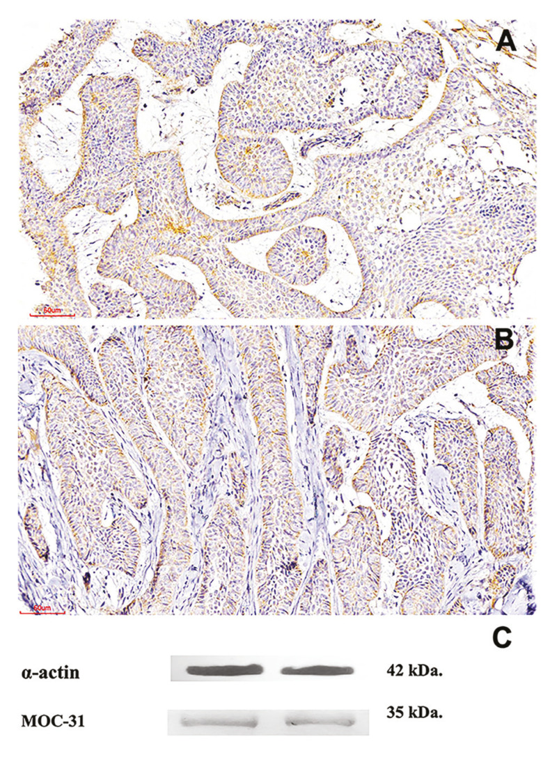
Ep-CAM (MOC-31) expression was evaluated by immunohistochemistry in tooth germs (TG) (n = 16) ameloblastomas (AM) (n = 60) and 2 ameloblastic carcinomas. Sections were visualized in their totality with an optical microscope, and positivity observed in cell membrane and cytoplasm was graded according to the following semi-quantitative scale: Neg, "essentially unstained", for negative sections or staining <5% of cells; + for staining of 5-50% of cells; ++ for staining >50% of cells.
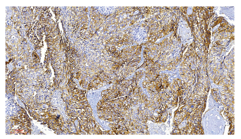
Most tooth germs expressed MOC-31 (81.3%), strong staining was observed both in the inner epithelium of the enamel organ and in the adjacent stellate reticulum. 16.7% of the AM cases showed MOC-31 expression, the immunoexpression expression was diffuse at the cytoplasmic and membrane level. The only two cases of ameloblastic carcinoma included were strong positive to MOC-31. No correlation was observed between protein expression and gender, age, clinical variants, or histological subtypes.
Overexpression was found in TG and ameloblastic carcinoma compared to AM; further studies with different experimental strategies are suggested to clarify the biological significance of this finding.
Ep-CAM 是一种在正常情况下表达于大多数上皮细胞的跨膜糖蛋白,在这些组织中具有多种功能,包括细胞黏附、增殖、分化、细胞周期调控、迁移和细胞内信号转导。它在大多数恶性肿瘤中也过度表达,参与肿瘤的起始、进展和转移扩散。该蛋白在口腔肿瘤中的表达和作用,特别是在牙源性肿瘤中的表达和作用尚未确定。本研究旨在分析 Ep-CAM 在牙源性肿瘤中的表达。
采用免疫组织化学法检测 16 例牙胚(TG)、60 例成釉细胞瘤(AM)和 2 例成釉细胞癌中 Ep-CAM(MOC-31)的表达。光学显微镜下观察组织切片的全貌,根据以下半定量评分标准评估细胞膜和细胞质的阳性程度:Neg,“基本无染色”,用于阴性切片或染色<5%的细胞;+,表示染色<50%的细胞;++,表示染色>50%的细胞。
大多数牙胚表达 MOC-31(81.3%),牙釉器内釉上皮和邻近的星网状层均有强烈染色。16.7%的 AM 病例表达 MOC-31,免疫组化染色在细胞质和细胞膜水平呈弥漫性。包括的 2 例成釉细胞癌均强烈阳性表达 MOC-31。蛋白表达与性别、年龄、临床变异型或组织学亚型之间无相关性。
与 AM 相比,TG 和成釉细胞癌中存在过度表达;建议采用不同的实验策略进一步研究,以阐明这一发现的生物学意义。